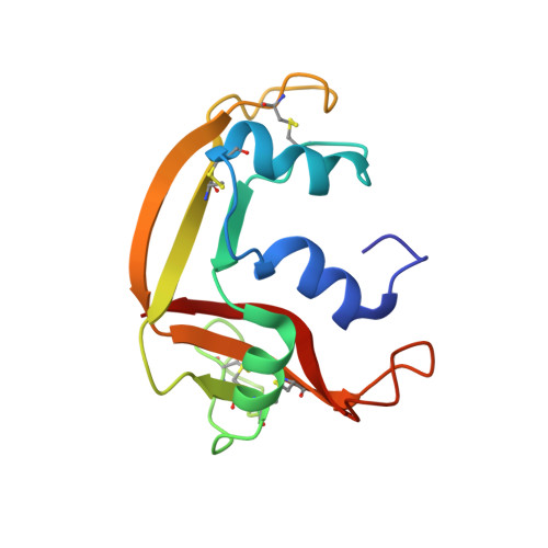
The Sulfate-Binding Site Structure of the Human Eosinophil Cationic Protein as Revealed by a New Crystal Form.
Boix, E., Pulido, D., Moussaoui, M., Nogues, V., Russi, S.(2012) J Struct Biol 179: 1
- PubMed: 22579681
- DOI: https://doi.org/10.1016/j.jsb.2012.04.023
- Primary Citation of Related Structures:
4A2O, 4A2Y - PubMed Abstract:
The human eosinophil cationic protein (ECP), also known as RNase 3, is an eosinophil secretion protein that is involved in innate immunity and displays antipathogen and proinflammatory activities. ECP has a high binding affinity for heterosaccharides, such as bacterial lipopolysaccharides and heparan sulfate found in the glycocalix of eukaryotic cells. We have crystallized ECP in complex with sulfate anions in a new monoclinic crystal form. In this form, the active site groove is exposed, providing an alternative model for ligand binding studies. By exploring the protein-sulfate complex, we have defined the sulfate binding site architecture. Three main sites (S1-S3) are located in the protein active site; S1 and S2 overlap with the phosphate binding sites involved in RNase nucleotide recognition. A new site (S3) that is unique to ECP is one of the key anchoring points for sulfated ligands. Arg 1 and Arg 7 in S3, together with Arg 34 and Arg 36 in S1, form the main basic clusters that assist in the recognition of ligand anionic groups. The location of additional sulfate bound molecules, some of which contribute to the crystal packing, may mimic the binding to extended anionic polymers. In conclusion, the structural data define a binding pattern for the recognition of sulfated molecules that can modulate the role of ECP in innate immunity. The results reveal the structural basis for the high affinity of ECP for glycosaminoglycans and can assist in structure-based drug design of inhibitors of the protein cytotoxicity to host tissues during inflammation.
Organizational Affiliation:
Department de Bioquímica i Biologia Molecular, Facultat de Biociències, Universitat Autònoma de Barcelona, Spain. Ester.Boix@uab.es