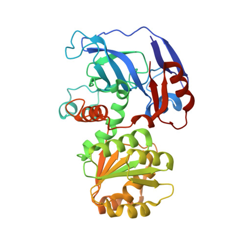
4A2C

Crystal structure of galactitol-1-phosphate dehydrogenase from Escherichia coli


Experimental Data Snapshot

  • Method: X-RAY DIFFRACTION
  • Resolution: 1.87 Å
  • R-Value Free: 0.273 
  • R-Value Work: 0.213 
  • R-Value Observed: 0.216 

wwPDB Validation   3D Report Full Report


This is version 1.3 of the entry. See complete history


Literature

The Crystal Structure of Galactitol-1-Phosphate 5-Dehydrogenase from Escherichia Coli K12 Provides Insights Into its Anomalous Behavior on Imac Processes

Esteban-Torres, M.Alvarez, Y.Acebron, I.De Las Rivas, B.Munoz, R.Kohring, G.-W.Roa, A.M.Sobrino, M.Mancheno, J.M.

(2012) FEBS Lett 586: 3127

  • DOI: https://doi.org/10.1016/j.febslet.2012.07.073
  • Primary Citation of Related Structures:  
    4A2C

  • PubMed Abstract: 

    Endogenous galactitol-1-phosphate 5-dehydrogenase (GPDH) (EC 1.1.1.251) from Escherichia coli spontaneously interacts with Ni(2+)-NTA matrices becoming a potential contaminant for recombinant, target His-tagged proteins. Purified recombinant, untagged GPDH (rGPDH) converted galactitol into tagatose, and d-tagatose-6-phosphate into galactitol-1-phosphate, in a Zn(2+)- and NAD(H)-dependent manner and readily crystallized what has permitted to solve its crystal structure. In contrast, N-terminally His-tagged GPDH was marginally stable and readily aggregated. The structure of rGPDH revealed metal-binding sites characteristic from the medium-chain dehydrogenase/reductase protein superfamily which may explain its ability to interact with immobilized metals. The structure also provides clues on the harmful effects of the N-terminal His-tag.


  • Organizational Affiliation

    Laboratorio de Biotecnología Bacteriana, Instituto Ciencia y Tecnología de Alimentos y Nutrición (ICTAN), CSIC, Madrid, Spain.


Macromolecules
Find similar proteins by:  (by identity cutoff)  |  3D Structure
Entity ID: 1
MoleculeChains Sequence LengthOrganismDetailsImage
GALACTITOL-1-PHOSPHATE 5-DEHYDROGENASE
A, B
346Escherichia coli K-12Mutation(s): 0 
EC: 1.1.1.251
UniProt
Find proteins for P0A9S3 (Escherichia coli (strain K12))
Explore P0A9S3 
Go to UniProtKB:  P0A9S3
Entity Groups  
Sequence Clusters30% Identity50% Identity70% Identity90% Identity95% Identity100% Identity
UniProt GroupP0A9S3
Sequence Annotations
Expand
  • Reference Sequence
Experimental Data & Validation

Experimental Data

  • Method: X-RAY DIFFRACTION
  • Resolution: 1.87 Å
  • R-Value Free: 0.273 
  • R-Value Work: 0.213 
  • R-Value Observed: 0.216 
  • Space Group: P 1 21 1
Unit Cell:
Length ( Å )Angle ( ˚ )
a = 43.72α = 90
b = 77.14β = 95.36
c = 107.51γ = 90
Software Package:
Software NamePurpose
PHENIXrefinement
MOSFLMdata reduction
SCALAdata scaling
PHASERphasing

Structure Validation

View Full Validation Report


Entry History 

Deposition Data

Revision History  (Full details and data files)

  • Version 1.0: 2012-08-08
    Type: Initial release
  • Version 1.1: 2012-08-22
    Changes: Database references
  • Version 1.2: 2012-10-03
    Changes: Database references
  • Version 1.3: 2023-12-20
    Changes: Data collection, Database references, Derived calculations, Other, Refinement description