3ZLZ

Lys6-linked tri-ubiquitin


Experimental Data Snapshot

  • Method: X-RAY DIFFRACTION
  • Resolution: 2.90 Å
  • R-Value Free: 0.288 
  • R-Value Work: 0.249 
  • R-Value Observed: 0.254 

wwPDB Validation   3D Report Full Report


This is version 1.4 of the entry. See complete history


Literature

Assembly, Analysis and Architecture of Atypical Ubiquitin Chains

Hospenthal, M.K.Freund, S.M.V.Komander, D.

(2013) Nat Struct Mol Biol 20: 555

  • DOI: https://doi.org/10.1038/nsmb.2547
  • Primary Citation of Related Structures:  
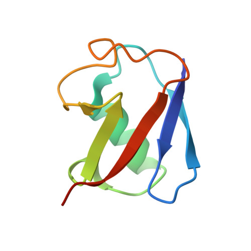
    3ZLZ

  • PubMed Abstract: 

    Ubiquitin (Ub) chains regulate many cellular processes, but several chain types including Lys6 linkages have remained unstudied. Here we analyze the bacterial effector E3 ligase NleL (non-Lee-encoded effector ligase) from enterohemorrhagic Escherichia coli (EHEC) O157:H7, which assembles Lys6- and Lys48-linked Ub polymers. Using linkage-specific human deubiquitinases (DUBs) we show that NleL generates heterotypic Ub chains, and branched chains are efficiently hydrolyzed by DUBs. USP family DUBs cleave Lys6-linked polymers exclusively from the distal end, whereas a DUB with preference for Lys6 can cleave Lys6-linked polymers at any position in the chain. We used NleL to generate large quantities of Lys6-linked polyUb. Crystallographic and NMR spectroscopy analyses revealed that an asymmetric interface between Ile44 and Ile36 hydrophobic patches of neighboring Ub moieties is propagated in longer Lys6-linked Ub chains. Interactions via the Ile36 patch can displace Leu8 from the Ile44 patch, leading to marked structural perturbations of Ub.


  • Organizational Affiliation

    Medical Research Council Laboratory of Molecular Biology, Cambridge, UK.


Macromolecules
Find similar proteins by:  (by identity cutoff)  |  3D Structure
Entity ID: 1
MoleculeChains Sequence LengthOrganismDetailsImage
UBIQUITIN
A, B
76Homo sapiensMutation(s): 1 
UniProt & NIH Common Fund Data Resources
Find proteins for P0CG47 (Homo sapiens)
Explore P0CG47 
Go to UniProtKB:  P0CG47
PHAROS:  P0CG47
GTEx:  ENSG00000170315 
Entity Groups  
Sequence Clusters30% Identity50% Identity70% Identity90% Identity95% Identity100% Identity
UniProt GroupP0CG47
Sequence Annotations
Expand
  • Reference Sequence
Experimental Data & Validation

Experimental Data

  • Method: X-RAY DIFFRACTION
  • Resolution: 2.90 Å
  • R-Value Free: 0.288 
  • R-Value Work: 0.249 
  • R-Value Observed: 0.254 
  • Space Group: P 43 3 2
Unit Cell:
Length ( Å )Angle ( ˚ )
a = 104.93α = 90
b = 104.93β = 90
c = 104.93γ = 90
Software Package:
Software NamePurpose
PHENIXrefinement
MOSFLMdata reduction
SCALAdata scaling
PHASERphasing

Structure Validation

View Full Validation Report


Entry History 

Deposition Data

Revision History  (Full details and data files)

  • Version 1.0: 2013-04-10
    Type: Initial release
  • Version 1.1: 2013-04-17
    Changes: Database references
  • Version 1.2: 2013-05-22
    Changes: Database references
  • Version 1.3: 2017-09-27
    Changes: Data collection
  • Version 1.4: 2023-12-20
    Changes: Data collection, Database references, Derived calculations, Other, Refinement description