


Crystal structure of Gox0525
Yuan, Y.A., Lin, J.P.To be published.
Experimental Data Snapshot
wwPDB Validation 3D Report Full Report

Entity ID: 1 | |||||
|---|---|---|---|---|---|
| Molecule | Chains | Sequence Length | Organism | Details | Image |
| Putative oxidoreductase | 246 | Gluconobacter oxydans 621H | Mutation(s): 0 Gene Names: GOX0525 EC: 1.1.1 | 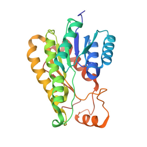 | |
UniProt | |||||
Find proteins for Q5FTJ3 (Gluconobacter oxydans (strain 621H)) Explore Q5FTJ3 Go to UniProtKB: Q5FTJ3 | |||||
Entity Groups | |||||
| Sequence Clusters | 30% Identity50% Identity70% Identity90% Identity95% Identity100% Identity | ||||
| UniProt Group | Q5FTJ3 | ||||
Sequence AnnotationsExpand | |||||
| |||||
| Length ( Å ) | Angle ( ˚ ) |
|---|---|
| a = 58.061 | α = 90 |
| b = 56.033 | β = 93.2 |
| c = 263.992 | γ = 90 |
| Software Name | Purpose |
|---|---|
| HKL-2000 | data collection |
| MOLREP | phasing |
| REFMAC | refinement |
| HKL-2000 | data reduction |
| HKL-2000 | data scaling |
RCSB PDB (citation) is hosted by
RCSB PDB is a member of the

