Structure of D-tagatose 3-epimerase-like protein from Methanocaldococcus jannaschii
Uechi, K., Takata, G., Yoneda, K., Ohshima, T., Sakuraba, H.(2014) Acta Crystallogr Sect F Struct Biol Cryst Commun 70: 890-895
- PubMed: 25005083
- DOI: https://doi.org/10.1107/S2053230X14011005
- Primary Citation of Related Structures:
3WQO - PubMed Abstract:
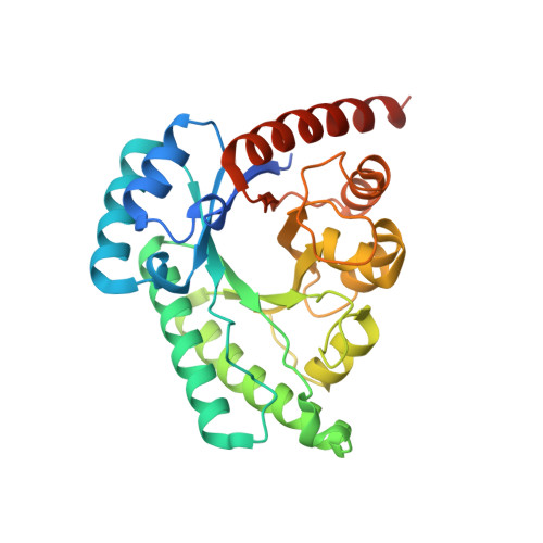
The crystal structure of a D-tagatose 3-epimerase-like protein (MJ1311p) encoded by a hypothetical open reading frame, MJ1311, in the genome of the hyperthermophilic archaeon Methanocaldococcus jannaschii was determined at a resolution of 2.64 Å. The asymmetric unit contained two homologous subunits, and the dimer was generated by twofold symmetry. The overall fold of the subunit proved to be similar to those of the D-tagatose 3-epimerase from Pseudomonas cichorii and the D-psicose 3-epimerases from Agrobacterium tumefaciens and Clostridium cellulolyticum. However, the situation at the subunit-subunit interface differed substantially from that in D-tagatose 3-epimerase family enzymes. In MJ1311p, Glu125, Leu126 and Trp127 from one subunit were found to be located over the metal-ion-binding site of the other subunit and appeared to contribute to the active site, narrowing the substrate-binding cleft. Moreover, the nine residues comprising a trinuclear zinc centre in endonuclease IV were found to be strictly conserved in MJ1311p, although a distinct groove involved in DNA binding was not present. These findings indicate that the active-site architecture of MJ1311p is quite unique and is substantially different from those of D-tagatose 3-epimerase family enzymes and endonuclease IV.
Organizational Affiliation:
Rare Sugar Research Center, Kagawa University, 2393 Ikenobe, Miki-cho, Kita-gun, Kagawa 761-0701, Japan.