3WPN

Kinesin spindle protein Eg5 in complex with ATP-competitive inhibitor PVZB1194


Experimental Data Snapshot

  • Method: X-RAY DIFFRACTION
  • Resolution: 2.80 Å
  • R-Value Free: 0.290 
  • R-Value Work: 0.225 
  • R-Value Observed: 0.231 

wwPDB Validation   3D Report Full Report


Ligand Structure Quality Assessment 


This is version 1.2 of the entry. See complete history


Literature

Structural basis of new allosteric inhibition in Kinesin spindle protein eg5

Yokoyama, H.Sawada, J.Katoh, S.Matsuno, K.Ogo, N.Ishikawa, Y.Hashimoto, H.Fujii, S.Asai, A.

(2015) ACS Chem Biol 10: 1128-1136

  • DOI: https://doi.org/10.1021/cb500939x
  • Primary Citation of Related Structures:  
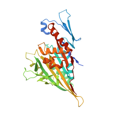
    3WPN

  • PubMed Abstract: 

    Kinesin spindle protein Eg5 is a target for anticancer therapies, and small molecule inhibitors of its ATPase activity have been developed. We herein report for the first time the crystal structure of and biochemical studies on the Eg5 motor domain in complex with a new type of allosteric inhibitor. The biphenyl-type inhibitor PVZB1194 binds to the α4/α6 allosteric pocket 15 Å from the ATP-binding pocket, which differs from conventional allosteric inhibitors that bind to the allosteric L5/α2/α3 pocket of Eg5. Binding of the inhibitor is involved in the neck-linker conformation and also causes conformational changes around the ATP-binding pocket through Tyr104 to affect the interaction of ATP with the pocket. This structure provides useful information for the development of novel types of allosteric drugs as well as a novel insight into the molecular mechanism responsible for regulating the motor activity of kinesins.


  • Organizational Affiliation

    †Department of Physical Biochemistry, School of Pharmaceutical Sciences, University of Shizuoka, Shizuoka 422-8526, Japan.


Macromolecules
Find similar proteins by:  (by identity cutoff)  |  3D Structure
Entity ID: 1
MoleculeChains Sequence LengthOrganismDetailsImage
Kinesin-like protein KIF11382Homo sapiensMutation(s): 0 
Gene Names: EG5KIF11
UniProt & NIH Common Fund Data Resources
Find proteins for P52732 (Homo sapiens)
Explore P52732 
Go to UniProtKB:  P52732
PHAROS:  P52732
GTEx:  ENSG00000138160 
Entity Groups  
Sequence Clusters30% Identity50% Identity70% Identity90% Identity95% Identity100% Identity
UniProt GroupP52732
Sequence Annotations
Expand
  • Reference Sequence
Small Molecules
Ligands 1 Unique
IDChains Name / Formula / InChI Key2D Diagram3D Interactions
B4S
Query on B4S

Download Ideal Coordinates CCD File 
B [auth A]3'-fluoro-4'-(trifluoromethyl)biphenyl-4-sulfonamide
C13 H9 F4 N O2 S
UEPKMFLFRNDVAW-UHFFFAOYSA-N
Binding Affinity Annotations 
IDSourceBinding Affinity
B4S BindingDB:  3WPN IC50: 120 (nM) from 1 assay(s)
Experimental Data & Validation

Experimental Data

  • Method: X-RAY DIFFRACTION
  • Resolution: 2.80 Å
  • R-Value Free: 0.290 
  • R-Value Work: 0.225 
  • R-Value Observed: 0.231 
  • Space Group: P 21 21 21
Unit Cell:
Length ( Å )Angle ( ˚ )
a = 71.912α = 90
b = 95.425β = 90
c = 59.015γ = 90
Software Package:
Software NamePurpose
HKL-2000data collection
MOLREPphasing
REFMACrefinement
HKL-2000data reduction
HKL-2000data scaling

Structure Validation

View Full Validation Report


Ligand Structure Quality Assessment 


Entry History 

Deposition Data

Revision History  (Full details and data files)

  • Version 1.0: 2015-01-21
    Type: Initial release
  • Version 1.1: 2015-04-29
    Changes: Database references
  • Version 1.2: 2023-11-08
    Changes: Data collection, Database references, Derived calculations, Refinement description