Structural fluctuation observed in Z-DNA d(CGCGCG)2 in the absence of divalent metal cations and polyamines
Chatake, T.(2013) J Synchrotron Radiat 20: 864-868
- PubMed: 24121329
- DOI: https://doi.org/10.1107/S0909049513020773
- Primary Citation of Related Structures:
3WBO - PubMed Abstract:
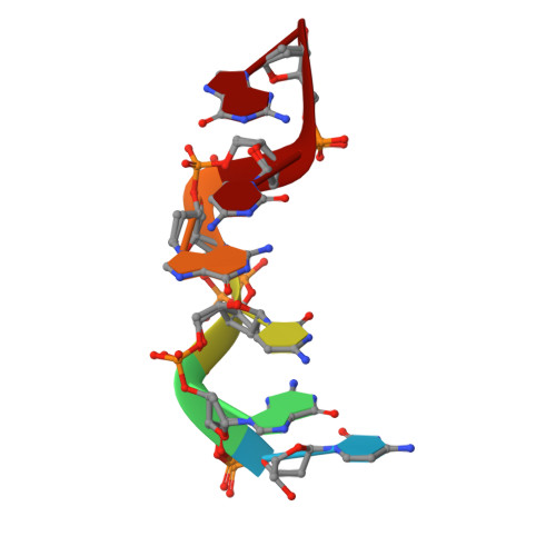
In the present study, Z-DNA d(CGCGCG)2 was crystallized from a DNA solution in the absence of divalent metal cations and polyamines, and its X-ray structure was determined at 0.98 Å resolution. Comparison of this structure and previously reported Z-DNA structures, containing Mg(2+) cations and/or polyamines, demonstrated that Z-DNA can have structural fluctuations with respect to phosphate groups and hydration in the minor groove. At the GpC steps, a two-state structural equilibrium between the ZI and ZII conformations was frequently observed. In contrast, at the CpG steps, the phosphate groups exhibited rotational fluctuation, which could induce distortion of sugar puckering. In addition, alternative positions of water molecules were found in the middle of the minor groove of the Z-DNA. These structural fluctuations were likely observable because of the absence of Mg(2+) cations and polyamines. The results related to these phenomena were supported by those of other experimental methods, suggesting the possibility of these fluctuations occurring in biological conditions.
Organizational Affiliation:
Research Reactor Institute, Kyoto University, Asashironishi 2, Kumatori, Osaka 590-0494, Japan.