


Crystal structure of unsaturated glucuronyl hydrolase mutant D115N from Streptcoccus agalactiae
Nakamichi, Y., Maruyama, Y., Mikami, B., Hashimoto, W., Murata, K.To be published.
Experimental Data Snapshot
wwPDB Validation 3D Report Full Report

Entity ID: 1 | |||||
|---|---|---|---|---|---|
| Molecule | Chains | Sequence Length | Organism | Details | Image |
| Putative uncharacterized protein gbs1889 | 398 | Streptococcus agalactiae NEM316 | Mutation(s): 1 Gene Names: gbs1889 EC: 3.2.1 | 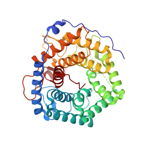 | |
UniProt | |||||
Find proteins for Q8E372 (Streptococcus agalactiae serotype III (strain NEM316)) Explore Q8E372 Go to UniProtKB: Q8E372 | |||||
Entity Groups | |||||
| Sequence Clusters | 30% Identity50% Identity70% Identity90% Identity95% Identity100% Identity | ||||
| UniProt Group | Q8E372 | ||||
Sequence AnnotationsExpand | |||||
| |||||
| Ligands 1 Unique | |||||
|---|---|---|---|---|---|
| ID | Chains | Name / Formula / InChI Key | 2D Diagram | 3D Interactions | |
| SO4 Query on SO4 | E [auth A] F [auth B] G [auth C] H [auth C] I [auth D] | SULFATE ION O4 S QAOWNCQODCNURD-UHFFFAOYSA-L |  | ||
| Length ( Å ) | Angle ( ˚ ) |
|---|---|
| a = 81.074 | α = 90 |
| b = 95.914 | β = 104.06 |
| c = 110.886 | γ = 90 |
| Software Name | Purpose |
|---|---|
| HKL-2000 | data collection |
| MOLREP | phasing |
| REFMAC | refinement |
| HKL-2000 | data reduction |
| HKL-2000 | data scaling |
RCSB PDB (citation) is hosted by
RCSB PDB is a member of the

