3VOH

CcCel6A catalytic domain complexed with cellobiose


Experimental Data Snapshot

  • Method: X-RAY DIFFRACTION
  • Resolution: 2.40 Å
  • R-Value Free: 0.246 
  • R-Value Work: 0.190 
  • R-Value Observed: 0.193 

wwPDB Validation   3D Report Full Report


Ligand Structure Quality Assessment 


This is version 2.1 of the entry. See complete history


Literature

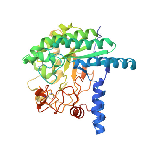
Comparison of the structural changes in two cellobiohydrolases, CcCel6A and CcCel6C, from Coprinopsis cinerea - a tweezer-like motion in the structure of CcCel6C

Tamura, M.Miyazaki, T.Tanaka, Y.Yoshida, M.Nishikawa, A.Tonozuka, T.

(2012) FEBS J 279: 1871-1882

  • DOI: https://doi.org/10.1111/j.1742-4658.2012.08568.x
  • Primary Citation of Related Structures:  
    3VOG, 3VOH, 3VOI, 3VOJ

  • PubMed Abstract: 

    The basidiomycete Coprinopsis cinerea produces five cellobiohydrolases belonging to glycoside hydrolase family 6 (GH6). Among these enzymes, C. cinerea cellulase 6C (CcCel6C), but not C. cinerea cellulase 6A (CcCel6A), can efficiently hydrolyze carboxymethyl cellulose and is constitutively expressed in C. cinerea. In contrast, CcCel6A possesses a cellulose-binding domain, and is strongly induced by cellobiose. Here, we determined the crystal structures of the CcCel6A catalytic domain complexed with a Hepes buffer molecule, with cellobiose, and with p-nitrophenyl β-D-cellotrioside (pNPG3). A notable feature of the GH6 cellobiohydrolases is that the active site is enclosed by two loops to form a tunnel, and the loops have been demonstrated to open and close in response to ligand binding. The enclosed tunnel of CcCel6A-Hepes is seen as the open form, whereas the tunnels of CcCel6A-cellobiose and CcCel6A-pNPG3 adopt the closed form. pNPG3 was not hydrolyzed by CcCel6A, and bound in subsites +1 to +4. On the basis of this observation, we constructed two mutants, CcCel6A D164A and CcCel6C D102A. Neither CcCel6A D164A nor CcCel6C D102A hydrolyze phosphoric acid-swollen cellulose. We have previously determined the crystal structures of CcCel6C unbound and in complex with ligand, both of which adopt the open form. In the present study, both CcCel6A and CcCel6C mutants were identified as the closed form. However, the motion angle of CcCel6C was more than 10-fold greater than that of CcCel6A. The width of the active site cleft of CcCel6C was narrowed, owing to a tweezer-like motion.


  • Organizational Affiliation

    Department of Applied Biological Science, Tokyo University of Agriculture and Technology, Tokyo, Japan.


Macromolecules
Find similar proteins by:  (by identity cutoff)  |  3D Structure
Entity ID: 1
MoleculeChains Sequence LengthOrganismDetailsImage
Cellobiohydrolase373Coprinopsis cinereaMutation(s): 0 
Gene Names: CcCel6A
EC: 3.2.1.91
UniProt
Find proteins for B7X9Z0 (Coprinopsis cinerea)
Explore B7X9Z0 
Go to UniProtKB:  B7X9Z0
Entity Groups  
Sequence Clusters30% Identity50% Identity70% Identity90% Identity95% Identity100% Identity
UniProt GroupB7X9Z0
Sequence Annotations
Expand
  • Reference Sequence
Oligosaccharides

Help

Entity ID: 2
MoleculeChains Length2D Diagram Glycosylation3D Interactions
beta-D-glucopyranose-(1-4)-beta-D-glucopyranose
B
2N/A
Glycosylation Resources
GlyTouCan:  G84824ZO
GlyCosmos:  G84824ZO
Entity ID: 3
MoleculeChains Length2D Diagram Glycosylation3D Interactions
beta-D-glucopyranose-(1-4)-beta-D-glucopyranose-(1-4)-beta-D-glucopyranose
C
3N/A
Glycosylation Resources
GlyTouCan:  G14338VK
GlyCosmos:  G14338VK
GlyGen:  G14338VK
Small Molecules
Ligands 1 Unique
IDChains Name / Formula / InChI Key2D Diagram3D Interactions
BGC
Query on BGC

Download Ideal Coordinates CCD File 
D [auth A]beta-D-glucopyranose
C6 H12 O6
WQZGKKKJIJFFOK-VFUOTHLCSA-N
Biologically Interesting Molecules (External Reference) 2 Unique
Experimental Data & Validation

Experimental Data

  • Method: X-RAY DIFFRACTION
  • Resolution: 2.40 Å
  • R-Value Free: 0.246 
  • R-Value Work: 0.190 
  • R-Value Observed: 0.193 
  • Space Group: P 21 21 21
Unit Cell:
Length ( Å )Angle ( ˚ )
a = 45.619α = 90
b = 72.14β = 90
c = 104.661γ = 90
Software Package:
Software NamePurpose
ADSCdata collection
MOLREPphasing
REFMACrefinement
HKL-2000data reduction
HKL-2000data scaling

Structure Validation

View Full Validation Report


Ligand Structure Quality Assessment 


Entry History 

Deposition Data

Revision History  (Full details and data files)

  • Version 1.0: 2012-03-21
    Type: Initial release
  • Version 1.1: 2012-05-02
    Changes: Database references
  • Version 2.0: 2020-07-29
    Type: Remediation
    Reason: Carbohydrate remediation
    Changes: Atomic model, Data collection, Database references, Derived calculations, Non-polymer description, Structure summary
  • Version 2.1: 2023-11-08
    Changes: Data collection, Database references, Refinement description, Structure summary