3VM5

Recombinant medaka fish alpha-amylase expressed in yeast Pichia pastoris


Experimental Data Snapshot

  • Method: X-RAY DIFFRACTION
  • Resolution: 2.85 Å
  • R-Value Free: 0.248 
  • R-Value Work: 0.177 
  • R-Value Observed: 0.180 

wwPDB Validation   3D Report Full Report


This is version 1.2 of the entry. See complete history


Literature

Structural and functional characterization of recombinant medaka fish alpha-amylase expressed in yeast Pichia pastoris.

Mizutani, K.Toyoda, M.Otake, Y.Yoshioka, S.Takahashi, N.Mikami, B.

(2012) Biochim Biophys Acta 1824: 954-962

  • DOI: https://doi.org/10.1016/j.bbapap.2012.05.005
  • Primary Citation of Related Structures:  
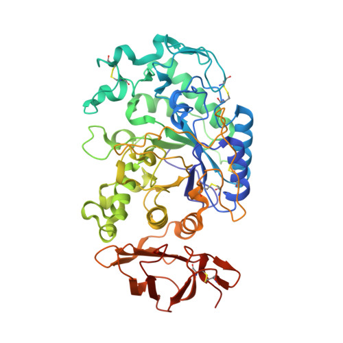
    3VM5

  • PubMed Abstract: 

    The medaka fish α-amylase was expressed and purified. The expression systems were constructed using methylotrophic yeast Pichia pastoris, and the recombinant proteins were secreted into the culture medium. Purified recombinant α-amylase exhibited starch hydrolysis activity. The optimal pH, denaturation temperature, and K(M) and V(max) values were determined; chloride ions were essential for enzyme activity. The purified protein was also crystallized and examined by X-ray crystallography. The structure has the (α/β)(8) barrel fold, as do other known α-amylases, and the overall structure is very similar to the structure of vertebrate (human and pig) α-amylases. A novel expression plasmid was developed. Using this plasmid, high-throughput construction of an expression system by homologous recombination in P. pastoris cells, previously reported for membrane proteins, was successfully applied to the secretory protein.


  • Organizational Affiliation

    Graduate School of Agriculture, Kyoto University, Kyoto, Japan. kmizutani@kais.kyoto-u.ac.jp


Macromolecules
Find similar proteins by:  (by identity cutoff)  |  3D Structure
Entity ID: 1
MoleculeChains Sequence LengthOrganismDetailsImage
alpha-amylase505Oryzias latipesMutation(s): 0 
UniProt
Find proteins for H2N0D4 (Oryzias latipes)
Explore H2N0D4 
Go to UniProtKB:  H2N0D4
Entity Groups  
Sequence Clusters30% Identity50% Identity70% Identity90% Identity95% Identity100% Identity
UniProt GroupH2N0D4
Sequence Annotations
Expand
  • Reference Sequence
Experimental Data & Validation

Experimental Data

  • Method: X-RAY DIFFRACTION
  • Resolution: 2.85 Å
  • R-Value Free: 0.248 
  • R-Value Work: 0.177 
  • R-Value Observed: 0.180 
  • Space Group: P 32 2 1
Unit Cell:
Length ( Å )Angle ( ˚ )
a = 83.045α = 90
b = 83.045β = 90
c = 174.228γ = 120
Software Package:
Software NamePurpose
Rayonixdata collection
PHASERphasing
PHENIXrefinement
HKL-2000data reduction
HKL-2000data scaling

Structure Validation

View Full Validation Report


Entry History 

Deposition Data

Revision History  (Full details and data files)

  • Version 1.0: 2012-06-06
    Type: Initial release
  • Version 1.1: 2013-06-26
    Changes: Database references
  • Version 1.2: 2023-11-08
    Changes: Data collection, Database references, Derived calculations, Refinement description