3V78

Crystal Structure of Transcriptional Regulator


Experimental Data Snapshot

  • Method: X-RAY DIFFRACTION
  • Resolution: 2.30 Å
  • R-Value Free: 0.262 
  • R-Value Work: 0.206 
  • R-Value Observed: 0.209 

wwPDB Validation   3D Report Full Report


Ligand Structure Quality Assessment 


This is version 1.2 of the entry. See complete history


Literature

Structural and functional analysis of the transcriptional regulator Rv3066 of Mycobacterium tuberculosis.

Bolla, J.R.Do, S.V.Long, F.Dai, L.Su, C.C.Lei, H.T.Chen, X.Gerkey, J.E.Murphy, D.C.Rajashankar, K.R.Zhang, Q.Yu, E.W.

(2012) Nucleic Acids Res 40: 9340-9355

  • DOI: https://doi.org/10.1093/nar/gks677
  • Primary Citation of Related Structures:  
    3V6G, 3V78

  • PubMed Abstract: 

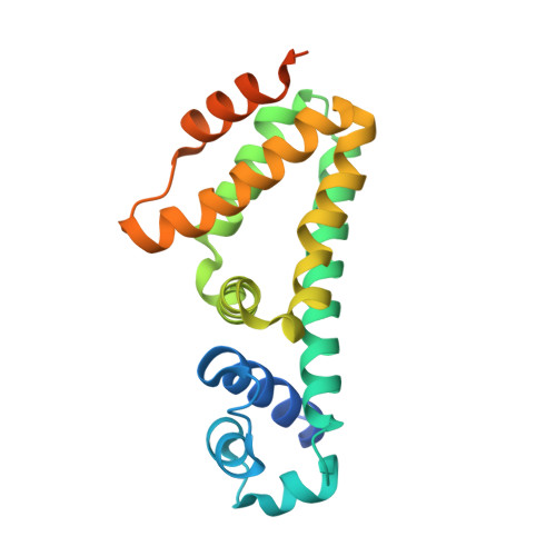
    The Mmr multidrug efflux pump recognizes and actively extrudes a broad range of antimicrobial agents, and promotes the intrinsic resistance to these antimicrobials in Mycobacterium tuberculosis. The expression of Mmr is controlled by the TetR-like transcriptional regulator Rv3066, whose open reading frame is located downstream of the mmr operon. To understand the structural basis of Rv3066 regulation, we have determined the crystal structures of Rv3066, both in the absence and presence of bound ethidium, revealing an asymmetric homodimeric two-domain molecule with an entirely helical architecture. The structures underscore the flexibility and plasticity of the regulator essential for multidrug recognition. Comparison of the apo-Rv3066 and Rv3066-ethidium crystal structures suggests that the conformational changes leading to drug-mediated derepression is primarily due to a rigid body rotational motion within the dimer interface of the regulator. The Rv3066 regulator creates a multidrug-binding pocket, which contains five aromatic residues. The bound ethidium is found buried within the multidrug-binding site, where extensive aromatic stacking interactions seemingly govern the binding. In vitro studies reveal that the dimeric Rv3066 regulator binds to a 14-bp palindromic inverted repeat sequence in the nanomolar range. These findings provide new insight into the mechanisms of ligand binding and Rv3066 regulation.


  • Organizational Affiliation

    Department of Chemistry, College of Veterinary Medicine, Iowa State University, Ames, IA 50011, USA.


Macromolecules
Find similar proteins by:  (by identity cutoff)  |  3D Structure
Entity ID: 1
MoleculeChains Sequence LengthOrganismDetailsImage
PROBABLE TRANSCRIPTIONAL REGULATORY PROTEIN (PROBABLY DEOR-FAMILY)
A, B
208Mycobacterium tuberculosisMutation(s): 0 
Gene Names: MT3151Rv3066
UniProt
Find proteins for P95092 (Mycobacterium tuberculosis (strain CDC 1551 / Oshkosh))
Explore P95092 
Go to UniProtKB:  P95092
Entity Groups  
Sequence Clusters30% Identity50% Identity70% Identity90% Identity95% Identity100% Identity
UniProt GroupP95092
Sequence Annotations
Expand
  • Reference Sequence
Small Molecules
Binding Affinity Annotations 
IDSourceBinding Affinity
ET Binding MOAD:  3V78 Kd: 2900 (nM) from 1 assay(s)
PDBBind:  3V78 Kd: 2900 (nM) from 1 assay(s)
Experimental Data & Validation

Experimental Data

  • Method: X-RAY DIFFRACTION
  • Resolution: 2.30 Å
  • R-Value Free: 0.262 
  • R-Value Work: 0.206 
  • R-Value Observed: 0.209 
  • Space Group: P 31 2 1
Unit Cell:
Length ( Å )Angle ( ˚ )
a = 99.098α = 90
b = 99.098β = 90
c = 66.478γ = 120
Software Package:
Software NamePurpose
PHENIXrefinement
SCALEPACKdata scaling
REFMACrefinement
PDB_EXTRACTdata extraction
ADSCdata collection
PHASERphasing
DENZOdata reduction

Structure Validation

View Full Validation Report


Ligand Structure Quality Assessment 


Entry History 

Deposition Data

Revision History  (Full details and data files)

  • Version 1.0: 2012-12-26
    Type: Initial release
  • Version 1.1: 2019-07-17
    Changes: Data collection, Refinement description
  • Version 1.2: 2023-09-13
    Changes: Data collection, Database references, Derived calculations, Refinement description