3UT0

Crystal structure of exo-1,3/1,4-beta-glucanase (EXOP) from Pseudoalteromonas sp. BB1


Experimental Data Snapshot

  • Method: X-RAY DIFFRACTION
  • Resolution: 2.30 Å
  • R-Value Free: 0.270 
  • R-Value Work: 0.223 
  • R-Value Observed: 0.225 

wwPDB Validation   3D Report Full Report


This is version 1.1 of the entry. See complete history


Literature

Structure and activity of exo-1,3/1,4-beta-glucanase from marine bacterium Pseudoalteromonas sp. BB1 showing a novel C-terminal domain

Nakatani, Y.Cutfield, S.M.Cowieson, N.P.Cutfield, J.F.

(2011) FEBS J 

  • DOI: https://doi.org/10.1111/j.1742-4658.2011.08439.x
  • Primary Citation of Related Structures:  
    3F95, 3RRX, 3USZ, 3UT0

  • PubMed Abstract: 

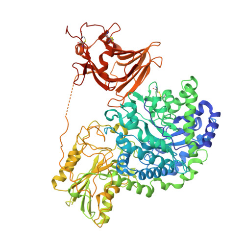
    Following the discovery of an exo-1,3/1,4-β-glucanase (glycoside hydrolase family 3) from a seaweed-associated bacterium Pseudoalteromonas sp. BB1, the recombinant three-domain protein (ExoP) was crystallized and its structure solved to 2.3 Å resolution. The first two domains of ExoP, both of which contribute to the architecture of the active site, are similar to those of the two-domain barley homologue β-d-glucan exohydrolase (ExoI) with a distinctive Trp-Trp clamp at the +1 subsite, although ExoI displays broader specificity towards β-glycosidic linkages. Notably, excision of the third domain of ExoP results in an inactive enzyme. Domain 3 has a β-sandwich structure and was shown by CD to be more temperature stable than the native enzyme. It makes relatively few contacts to domain 1 and none at all to domain 2. Two of the domain 3 residues involved at the interface, Q683 (forming one hydrogen bond) and Q676 (forming two hydrogen bonds) were mutated to alanine. Variant Q676A retained about half the activity of native ExoP, but the Q683A variant was severely attenuated. The crystal structure of Q683A-ExoP indicated that domain 3 was highly mobile and that Q683 is critical to the stabilization of ExoP by domain 3. Small-angle X-ray scattering data lent support to this proposal. Domain 3 does not appear to be an obvious carbohydrate-binding domain and is related neither in sequence nor structure to the additional domains characterized in other glycoside hydrolase 3 subgroups. Its major role appears to be for protein stability but it may also help orient substrate.


  • Organizational Affiliation

    Department of Biochemistry, University of Otago, Dunedin, New Zealand.


Macromolecules
Find similar proteins by:  (by identity cutoff)  |  3D Structure
Entity ID: 1
MoleculeChains Sequence LengthOrganismDetailsImage
Exo-1,3/1,4-beta-glucanase
A, B, C, D
822Pseudoalteromonas sp. BB1Mutation(s): 0 
Gene Names: exoP
EC: 3.2.1
UniProt
Find proteins for Q0QJA3 (Pseudoalteromonas sp. BB1)
Explore Q0QJA3 
Go to UniProtKB:  Q0QJA3
Entity Groups  
Sequence Clusters30% Identity50% Identity70% Identity90% Identity95% Identity100% Identity
UniProt GroupQ0QJA3
Sequence Annotations
Expand
  • Reference Sequence
Small Molecules
Ligands 4 Unique
IDChains Name / Formula / InChI Key2D Diagram3D Interactions
SO4
Query on SO4

Download Ideal Coordinates CCD File 
AA [auth C]
BA [auth C]
CA [auth C]
DA [auth C]
G [auth A]
AA [auth C],
BA [auth C],
CA [auth C],
DA [auth C],
G [auth A],
H [auth A],
I [auth A],
IA [auth D],
J [auth A],
JA [auth D],
K [auth A],
KA [auth D],
Q [auth B],
R [auth B],
S [auth B],
T [auth B],
U [auth B],
Z [auth C]
SULFATE ION
O4 S
QAOWNCQODCNURD-UHFFFAOYSA-L
EDO
Query on EDO

Download Ideal Coordinates CCD File 
EA [auth C]
FA [auth C]
L [auth A]
LA [auth D]
M [auth A]
EA [auth C],
FA [auth C],
L [auth A],
LA [auth D],
M [auth A],
MA [auth D],
N [auth A],
V [auth B],
W [auth B]
1,2-ETHANEDIOL
C2 H6 O2
LYCAIKOWRPUZTN-UHFFFAOYSA-N
CA
Query on CA

Download Ideal Coordinates CCD File 
E [auth A],
GA [auth D],
O [auth B],
X [auth C]
CALCIUM ION
Ca
BHPQYMZQTOCNFJ-UHFFFAOYSA-N
NA
Query on NA

Download Ideal Coordinates CCD File 
F [auth A],
HA [auth D],
P [auth B],
Y [auth C]
SODIUM ION
Na
FKNQFGJONOIPTF-UHFFFAOYSA-N
Experimental Data & Validation

Experimental Data

  • Method: X-RAY DIFFRACTION
  • Resolution: 2.30 Å
  • R-Value Free: 0.270 
  • R-Value Work: 0.223 
  • R-Value Observed: 0.225 
  • Space Group: P 1 21 1
Unit Cell:
Length ( Å )Angle ( ˚ )
a = 84.908α = 90
b = 257.821β = 115.47
c = 85.605γ = 90
Software Package:
Software NamePurpose
Blu-Icedata collection
PHASERphasing
PHENIXrefinement
XDSdata reduction
SCALAdata scaling

Structure Validation

View Full Validation Report


Entry History 

Deposition Data

Revision History  (Full details and data files)

  • Version 1.0: 2011-12-21
    Type: Initial release
  • Version 1.1: 2023-11-08
    Changes: Data collection, Database references, Derived calculations, Refinement description