3UGF

Crystal structure of a 6-SST/6-SFT from Pachysandra terminalis


Experimental Data Snapshot

  • Method: X-RAY DIFFRACTION
  • Resolution: 1.70 Å
  • R-Value Free: 0.186 
  • R-Value Work: 0.171 
  • R-Value Observed: 0.172 

wwPDB Validation   3D Report Full Report


This is version 2.1 of the entry. See complete history


Literature

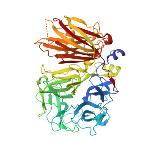
Crystal structure of 6-SST/6-SFT from Pachysandra terminalis, a plant fructan biosynthesizing enzyme in complex with its acceptor substrate 6-kestose.

Lammens, W.Le Roy, K.Yuan, S.Vergauwen, R.Rabijns, A.Van Laere, A.Strelkov, S.V.Van den Ende, W.

(2012) Plant J 70: 205-219

  • DOI: https://doi.org/10.1111/j.1365-313X.2011.04858.x
  • Primary Citation of Related Structures:  
    3UGF, 3UGG, 3UGH

  • PubMed Abstract: 

    Fructans play important roles as reserve carbohydrates and stress protectants in plants, and additionally serve as prebiotics with emerging antioxidant properties. Various fructan types are synthesized by an array of plant fructosyltransferases belonging to family 32 of the glycoside hydrolases (GH32), clustering together with GH68 in Clan-J. Here, the 3D structure of a plant fructosyltransferase from a native source, the Pachysandra terminalis 6-SST/6-SFT (Pt6-SST/6-SFT), is reported. In addition to its 1-SST (1-kestose-forming) and hydrolytic side activities, the enzyme uses sucrose to create graminan- and levan-type fructans, which are probably associated with cold tolerance in this species. Furthermore, a Pt6-SST/6-SFT complex with 6-kestose was generated, representing a genuine acceptor binding modus at the +1, +2 and +3 subsites in the active site. The enzyme shows a unique configuration in the vicinity of its active site, including a unique D/Q couple located at the +1 subsite that plays a dual role in donor and acceptor substrate binding. Furthermore, it shows a unique orientation of some hydrophobic residues, probably contributing to its specific functionality. A model is presented showing formation of a β(2-6) fructosyl linkage on 6-kestose to create 6,6-nystose, a mechanism that differs from the creation of a β(2-1) fructosyl linkage on sucrose to produce 1-kestose. The structures shed light on the evolution of plant fructosyltransferases from their vacuolar invertase ancestors, and contribute to further understanding of the complex structure-function relationships within plant GH32 members.


  • Organizational Affiliation

    Biology Department, Laboratory for Molecular Plant Physiology, Katholieke Universiteit Leuven, Kasteelpark Arenberg 31, Box 2434, B-3001 Heverlee, Belgium.


Macromolecules
Find similar proteins by:  (by identity cutoff)  |  3D Structure
Entity ID: 1
MoleculeChains Sequence LengthOrganismDetailsImage
Sucrose:(Sucrose/fructan) 6-fructosyltransferase
A, B
546Pachysandra terminalisMutation(s): 0 
UniProt
Find proteins for E3PQS3 (Pachysandra terminalis)
Explore E3PQS3 
Go to UniProtKB:  E3PQS3
Entity Groups  
Sequence Clusters30% Identity50% Identity70% Identity90% Identity95% Identity100% Identity
UniProt GroupE3PQS3
Sequence Annotations
Expand
  • Reference Sequence
Oligosaccharides

Help

Entity ID: 2
MoleculeChains Length2D Diagram Glycosylation3D Interactions
alpha-L-fucopyranose-(1-3)-2-acetamido-2-deoxy-beta-D-glucopyranose
C
2N-Glycosylation
Glycosylation Resources
GlyTouCan:  G80587NA
GlyCosmos:  G80587NA
GlyGen:  G80587NA
Entity ID: 3
MoleculeChains Length2D Diagram Glycosylation3D Interactions
alpha-L-fucopyranose-(1-3)-[2-acetamido-2-deoxy-beta-D-glucopyranose-(1-4)]2-acetamido-2-deoxy-beta-D-glucopyranose
D, H
3N-Glycosylation
Glycosylation Resources
GlyTouCan:  G11971MR
GlyCosmos:  G11971MR
GlyGen:  G11971MR
Entity ID: 4
MoleculeChains Length2D Diagram Glycosylation3D Interactions
beta-D-mannopyranose-(1-4)-2-acetamido-2-deoxy-beta-D-glucopyranose-(1-4)-2-acetamido-2-deoxy-beta-D-glucopyranose
E, G
3N-Glycosylation
Glycosylation Resources
GlyTouCan:  G15407YE
GlyCosmos:  G15407YE
GlyGen:  G15407YE
Entity ID: 5
MoleculeChains Length2D Diagram Glycosylation3D Interactions
beta-D-mannopyranose-(1-4)-2-acetamido-2-deoxy-beta-D-glucopyranose-(1-4)-[alpha-L-fucopyranose-(1-3)]2-acetamido-2-deoxy-beta-D-glucopyranose
F
4N-Glycosylation
Glycosylation Resources
GlyTouCan:  G18638YB
GlyCosmos:  G18638YB
GlyGen:  G18638YB
Entity ID: 6
MoleculeChains Length2D Diagram Glycosylation3D Interactions
alpha-D-mannopyranose-(1-6)-beta-D-mannopyranose-(1-4)-2-acetamido-2-deoxy-beta-D-glucopyranose-(1-4)-[alpha-L-fucopyranose-(1-3)]2-acetamido-2-deoxy-beta-D-glucopyranose
I
5N-Glycosylation
Glycosylation Resources
GlyTouCan:  G83878UZ
GlyCosmos:  G83878UZ
GlyGen:  G83878UZ
Entity ID: 7
MoleculeChains Length2D Diagram Glycosylation3D Interactions
2-acetamido-2-deoxy-beta-D-glucopyranose-(1-2)-alpha-D-mannopyranose-(1-3)-beta-D-mannopyranose-(1-4)-2-acetamido-2-deoxy-beta-D-glucopyranose-(1-4)-[alpha-L-fucopyranose-(1-3)]2-acetamido-2-deoxy-beta-D-glucopyranose
J
6N-Glycosylation
Glycosylation Resources
GlyTouCan:  G68807DB
GlyCosmos:  G68807DB
GlyGen:  G68807DB
Small Molecules
Ligands 2 Unique
IDChains Name / Formula / InChI Key2D Diagram3D Interactions
SO4
Query on SO4

Download Ideal Coordinates CCD File 
Q [auth A],
Y [auth B]
SULFATE ION
O4 S
QAOWNCQODCNURD-UHFFFAOYSA-L
GOL
Query on GOL

Download Ideal Coordinates CCD File 
K [auth A]
L [auth A]
M [auth A]
N [auth A]
O [auth A]
K [auth A],
L [auth A],
M [auth A],
N [auth A],
O [auth A],
P [auth A],
R [auth B],
S [auth B],
T [auth B],
U [auth B],
V [auth B],
W [auth B],
X [auth B]
GLYCEROL
C3 H8 O3
PEDCQBHIVMGVHV-UHFFFAOYSA-N
Experimental Data & Validation

Experimental Data

  • Method: X-RAY DIFFRACTION
  • Resolution: 1.70 Å
  • R-Value Free: 0.186 
  • R-Value Work: 0.171 
  • R-Value Observed: 0.172 
  • Space Group: P 21 21 21
Unit Cell:
Length ( Å )Angle ( ˚ )
a = 89.08α = 90
b = 125.27β = 90
c = 148.95γ = 90
Software Package:
Software NamePurpose
SCALAdata scaling
REFMACrefinement
PDB_EXTRACTdata extraction
HKL-2000data collection
XDSdata reduction
MOLREPphasing

Structure Validation

View Full Validation Report


Entry History 

Deposition Data

Revision History  (Full details and data files)

  • Version 1.0: 2011-11-30
    Type: Initial release
  • Version 1.1: 2012-10-24
    Changes: Database references
  • Version 2.0: 2020-07-29
    Type: Remediation
    Reason: Carbohydrate remediation
    Changes: Atomic model, Data collection, Derived calculations, Structure summary
  • Version 2.1: 2023-09-13
    Changes: Data collection, Database references, Refinement description, Structure summary