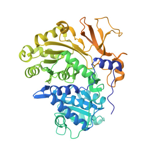
3U16

Structure of BasE N-terminal domain from Acinetobacter baumannii bound to 6-(p-benzyloxy)phenyl-1-(pyridin-4-ylmethyl)-1H-pyrazolo[3,4-b]pyridine-4-carboxylic acid.


Experimental Data Snapshot

  • Method: X-RAY DIFFRACTION
  • Resolution: 2.10 Å
  • R-Value Free: 0.219 
  • R-Value Work: 0.185 
  • R-Value Observed: 0.187 

wwPDB Validation   3D Report Full Report


Ligand Structure Quality Assessment 


This is version 1.3 of the entry. See complete history


Literature

Non-nucleoside inhibitors of BasE, an adenylating enzyme in the siderophore biosynthetic pathway of the opportunistic pathogen Acinetobacter baumannii.

Neres, J.Engelhart, C.A.Drake, E.J.Wilson, D.J.Fu, P.Boshoff, H.I.Barry 3rd, C.E.Gulick, A.M.Aldrich, C.C.

(2013) J Med Chem 56: 2385-2405

  • DOI: https://doi.org/10.1021/jm301709s
  • Primary Citation of Related Structures:  
    3U16, 3U17

  • PubMed Abstract: 

    Siderophores are small-molecule iron chelators produced by bacteria and other microorganisms for survival under iron limiting conditions such as found in a mammalian host. Siderophore biosynthesis is essential for the virulence of many important Gram-negative pathogens including Acinetobacter baumannii, Klebsiella pneumoniae, Pseudomonas aeruginosa, and Escherichia coli. We performed high-throughput screening against BasE, which is involved in siderophore biosynthesis in A. baumannii, and identified 6-phenyl-1-(pyridin-4-ylmethyl)-1H-pyrazolo[3,4-b]pyridine-4-carboxylic acid 15. Herein we report the synthesis, biochemical, and microbiological evaluation of a systematic series of analogues of the HTS hit 15. Analogue 67 is the most potent analogue with a KD of 2 nM against BasE. Structural characterization of the inhibitors with BasE reveals that they bind in a unique orientation in the active site, occupying all three substrate binding sites, and thus can be considered as multisubstrate inhibitors. These results provide a foundation for future studies aimed at increasing both enzyme potency and antibacterial activity.


  • Organizational Affiliation

    Center for Drug Design, Academic Health Center, University of Minnesota , Minneapolis, Minnesota 55455, USA.


Macromolecules
Find similar proteins by:  (by identity cutoff)  |  3D Structure
Entity ID: 1
MoleculeChains Sequence LengthOrganismDetailsImage
Peptide arylation enzyme
A, B
544Acinetobacter baumannii ACICUMutation(s): 0 
Gene Names: ACICU_02578basE
EC: 6.2.1
Entity Groups  
Sequence Clusters30% Identity50% Identity70% Identity90% Identity95% Identity100% Identity
Sequence Annotations
Expand
  • Reference Sequence
Small Molecules
Ligands 4 Unique
IDChains Name / Formula / InChI Key2D Diagram3D Interactions
H89
Query on H89

Download Ideal Coordinates CCD File 
C [auth A],
I [auth B]
6-[4-(benzyloxy)phenyl]-1-(pyridin-4-ylmethyl)-1H-pyrazolo[3,4-b]pyridine-4-carboxylic acid
C26 H20 N4 O3
LEKPQTJLNDALBT-UHFFFAOYSA-N
MRD
Query on MRD

Download Ideal Coordinates CCD File 
L [auth B](4R)-2-METHYLPENTANE-2,4-DIOL
C6 H14 O2
SVTBMSDMJJWYQN-RXMQYKEDSA-N
MPD
Query on MPD

Download Ideal Coordinates CCD File 
H [auth A](4S)-2-METHYL-2,4-PENTANEDIOL
C6 H14 O2
SVTBMSDMJJWYQN-YFKPBYRVSA-N
CA
Query on CA

Download Ideal Coordinates CCD File 
D [auth A]
E [auth A]
F [auth A]
G [auth A]
J [auth B]
D [auth A],
E [auth A],
F [auth A],
G [auth A],
J [auth B],
K [auth B]
CALCIUM ION
Ca
BHPQYMZQTOCNFJ-UHFFFAOYSA-N
Experimental Data & Validation

Experimental Data

  • Method: X-RAY DIFFRACTION
  • Resolution: 2.10 Å
  • R-Value Free: 0.219 
  • R-Value Work: 0.185 
  • R-Value Observed: 0.187 
  • Space Group: P 21 21 21
Unit Cell:
Length ( Å )Angle ( ˚ )
a = 66.093α = 90
b = 144.78β = 90
c = 148.675γ = 90
Software Package:
Software NamePurpose
PHENIXrefinement
REFMACrefinement
Blu-Icedata collection
HKL-2000data reduction
HKL-2000data scaling
REFMACphasing

Structure Validation

View Full Validation Report


Ligand Structure Quality Assessment 


Entry History 

Deposition Data

Revision History  (Full details and data files)

  • Version 1.0: 2012-10-03
    Type: Initial release
  • Version 1.1: 2019-07-17
    Changes: Data collection, Refinement description
  • Version 1.2: 2020-05-13
    Changes: Database references
  • Version 1.3: 2023-09-13
    Changes: Data collection, Database references, Derived calculations, Refinement description