3SYR

Glycogen phosphorylase b in complex with beta-D-glucopyranonucleoside 5-fluorouracil


Experimental Data Snapshot

  • Method: X-RAY DIFFRACTION
  • Resolution: 2.40 Å
  • R-Value Free: 0.217 
  • R-Value Work: 0.176 
  • R-Value Observed: 0.178 

wwPDB Validation   3D Report Full Report


Ligand Structure Quality Assessment 


This is version 2.0 of the entry. See complete history


Literature

3'-Axial CH(2) OH Substitution on Glucopyranose does not Increase Glycogen Phosphorylase Inhibitory Potency. QM/MM-PBSA Calculations Suggest Why.

Manta, S.Xipnitou, A.Kiritsis, C.Kantsadi, A.L.Hayes, J.M.Skamnaki, V.T.Lamprakis, C.Kontou, M.Zoumpoulakis, P.Zographos, S.E.Leonidas, D.D.Komiotis, D.

(2012) Chem Biol Drug Des 79: 663-673

  • DOI: https://doi.org/10.1111/j.1747-0285.2012.01349.x
  • Primary Citation of Related Structures:  
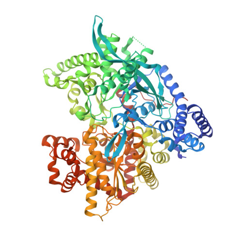
    3SYM, 3SYR

  • PubMed Abstract: 

    Glycogen phosphorylase is a molecular target for the design of potential hypoglycemic agents. Structure-based design pinpointed that the 3'-position of glucopyranose equipped with a suitable group has the potential to form interactions with enzyme's cofactor, pyridoxal 5'-phosphate (PLP), thus enhancing the inhibitory potency. Hence, we have investigated the binding of two ligands, 1-(β-d-glucopyranosyl)5-fluorouracil (GlcFU) and its 3'-CH(2) OH glucopyranose derivative. Both ligands were found to be low micromolar inhibitors with K(i) values of 7.9 and 27.1 μm, respectively. X-ray crystallography revealed that the 3'-CH(2) OH glucopyranose substituent is indeed involved in additional molecular interactions with the PLP γ-phosphate compared with GlcFU. However, it is 3.4 times less potent. To elucidate this discovery, docking followed by postdocking Quantum Mechanics/Molecular Mechanics - Poisson-Boltzmann Surface Area (QM/MM-PBSA) binding affinity calculations were performed. While the docking predictions failed to reflect the kinetic results, the QM/MM-PBSA revealed that the desolvation energy cost for binding of the 3'-CH(2) OH-substituted glucopyranose derivative out-weigh the enthalpy gains from the extra contacts formed. The benefits of performing postdocking calculations employing a more accurate solvation model and the QM/MM-PBSA methodology in lead optimization are therefore highlighted, specifically when the role of a highly polar/charged binding interface is significant.


  • Organizational Affiliation

    Department of Biochemistry and Biotechnology, University of Thessaly, 26 Ploutonos Str., Larissa, Greece.


Macromolecules
Find similar proteins by:  (by identity cutoff)  |  3D Structure
Entity ID: 1
MoleculeChains Sequence LengthOrganismDetailsImage
Glycogen phosphorylase, muscle form842Oryctolagus cuniculusMutation(s): 0 
EC: 2.4.1.1
UniProt
Find proteins for P00489 (Oryctolagus cuniculus)
Explore P00489 
Go to UniProtKB:  P00489
Entity Groups  
Sequence Clusters30% Identity50% Identity70% Identity90% Identity95% Identity100% Identity
UniProt GroupP00489
Sequence Annotations
Expand
  • Reference Sequence
Small Molecules
Ligands 1 Unique
IDChains Name / Formula / InChI Key2D Diagram3D Interactions
GPK
Query on GPK

Download Ideal Coordinates CCD File 
B [auth A]5-fluoro-1-(beta-D-glucopyranosyl)pyrimidine-2,4(1H,3H)-dione
C10 H13 F N2 O7
YHWGRVDTEORPPJ-XSEHCYKFSA-N
Modified Residues  1 Unique
IDChains TypeFormula2D DiagramParent
LLP
Query on LLP
A
L-PEPTIDE LINKINGC14 H22 N3 O7 PLYS
Binding Affinity Annotations 
IDSourceBinding Affinity
GPK BindingDB:  3SYR Ki: 5500 (nM) from 1 assay(s)
PDBBind:  3SYR Ki: 7900 (nM) from 1 assay(s)
Binding MOAD:  3SYR Ki: 7900 (nM) from 1 assay(s)
Experimental Data & Validation

Experimental Data

  • Method: X-RAY DIFFRACTION
  • Resolution: 2.40 Å
  • R-Value Free: 0.217 
  • R-Value Work: 0.176 
  • R-Value Observed: 0.178 
  • Space Group: P 43 21 2
Unit Cell:
Length ( Å )Angle ( ˚ )
a = 128.556α = 90
b = 128.556β = 90
c = 116.282γ = 90
Software Package:
Software NamePurpose
CrysalisProdata collection
REFMACrefinement
MOSFLMdata reduction
SCALAdata scaling
REFMACphasing

Structure Validation

View Full Validation Report


Ligand Structure Quality Assessment 


Entry History 

Deposition Data

Revision History  (Full details and data files)

  • Version 1.0: 2012-02-15
    Type: Initial release
  • Version 1.1: 2012-04-18
    Changes: Database references
  • Version 1.2: 2017-11-08
    Changes: Refinement description
  • Version 2.0: 2020-07-29
    Type: Remediation
    Reason: Carbohydrate remediation
    Changes: Atomic model, Data collection, Derived calculations