3SU9

E. Cloacae MURA in complex with UDP-N-acetylmuramic acid and covalent adduct of PEP with Cys115


Experimental Data Snapshot

  • Method: X-RAY DIFFRACTION
  • Resolution: 2.20 Å
  • R-Value Free: 0.210 
  • R-Value Work: 0.162 
  • R-Value Observed: 0.162 

wwPDB Validation   3D Report Full Report


Ligand Structure Quality Assessment 


This is version 1.4 of the entry. See complete history


Literature

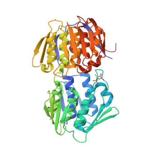
Functional Consequence of Covalent Reaction of Phosphoenolpyruvate with UDP-N-acetylglucosamine 1-Carboxyvinyltransferase (MurA).

Zhu, J.Y.Yang, Y.Han, H.Betzi, S.Olesen, S.H.Marsilio, F.Schonbrunn, E.

(2012) J Biol Chem 287: 12657-12667

  • DOI: https://doi.org/10.1074/jbc.M112.342725
  • Primary Citation of Related Structures:  
    3SPB, 3SU9, 3SWA, 3SWD, 3SWE, 3SWG, 3SWI, 3SWQ, 3UPK, 3V4T, 3V5V

  • PubMed Abstract: 

    The enzyme MurA has been an established antibiotic target since the discovery of fosfomycin, which specifically inhibits MurA by covalent modification of the active site residue Cys-115. Early biochemical studies established that Cys-115 also covalently reacts with substrate phosphoenolpyruvate (PEP) to yield a phospholactoyl adduct, but the structural and functional consequences of this reaction remained obscure. We captured and depicted the Cys-115-PEP adduct of Enterobacter cloacae MurA in various reaction states by X-ray crystallography. The data suggest that cellular MurA predominantly exists in a tightly locked complex with UDP-N-acetylmuramic acid (UNAM), the product of the MurB reaction, with PEP covalently attached to Cys-115. The uniqueness and rigidity of this "dormant" complex was previously not recognized and presumably accounts for the failure of drug discovery efforts toward the identification of novel and effective MurA inhibitors. We demonstrate that recently published crystal structures of MurA from various organisms determined by different laboratories were indeed misinterpreted and actually contain UNAM and covalently bound PEP. The Cys-115-PEP adduct was also captured in vitro during the reaction of free MurA and substrate UDP-N-acetylglucosamine or isomer UDP-N-acetylgalactosamine. The now available series of crystal structures allows a comprehensive view of the reaction cycle of MurA. It appears that the covalent reaction of MurA with PEP fulfills dual functions by tightening the complex with UNAM for the efficient feedback regulation of murein biosynthesis and by priming the PEP molecule for instantaneous reaction with substrate UDP-N-acetylglucosamine.


  • Organizational Affiliation

    Drug Discovery Department, Moffitt Cancer Center and Research Institute, Tampa, Florida 33612, USA.


Macromolecules
Find similar proteins by:  (by identity cutoff)  |  3D Structure
Entity ID: 1
MoleculeChains Sequence LengthOrganismDetailsImage
UDP-N-acetylglucosamine 1-carboxyvinyltransferase419Enterobacter cloacae subsp. cloacae ATCC 13047Mutation(s): 0 
Gene Names: murAmurZECL_04571
EC: 2.5.1.7
UniProt
Find proteins for P33038 (Enterobacter cloacae subsp. cloacae (strain ATCC 13047 / DSM 30054 / NBRC 13535 / NCTC 10005 / WDCM 00083 / NCDC 279-56))
Explore P33038 
Go to UniProtKB:  P33038
Entity Groups  
Sequence Clusters30% Identity50% Identity70% Identity90% Identity95% Identity100% Identity
UniProt GroupP33038
Sequence Annotations
Expand
  • Reference Sequence
Small Molecules
Ligands 3 Unique
IDChains Name / Formula / InChI Key2D Diagram3D Interactions
EPZ
Query on EPZ

Download Ideal Coordinates CCD File 
I [auth A](2R)-2-{[(2R,3R,4R,5S,6R)-3-(acetylamino)-2-{[(S)-{[(R)-{[(2R,3S,4R,5R)-5-(2,4-dioxo-3,4-dihydropyrimidin-1(2H)-yl)-3,4-dihydroxytetrahydrofuran-2-yl]methoxy}(hydroxy)phosphoryl]oxy}(hydroxy)phosphoryl]oxy}-5-hydroxy-6-(hydroxymethyl)tetrahydro-2H-pyran-4-yl]oxy}propanoic acid
C20 H31 N3 O19 P2
NQBRVZNDBBMBLJ-MQTLHLSBSA-N
EDO
Query on EDO

Download Ideal Coordinates CCD File 
B [auth A]
C [auth A]
D [auth A]
E [auth A]
F [auth A]
B [auth A],
C [auth A],
D [auth A],
E [auth A],
F [auth A],
G [auth A]
1,2-ETHANEDIOL
C2 H6 O2
LYCAIKOWRPUZTN-UHFFFAOYSA-N
ACT
Query on ACT

Download Ideal Coordinates CCD File 
H [auth A]ACETATE ION
C2 H3 O2
QTBSBXVTEAMEQO-UHFFFAOYSA-M
Modified Residues  1 Unique
IDChains TypeFormula2D DiagramParent
QPA
Query on QPA
A
L-PEPTIDE LINKINGC6 H12 N O8 P SCYS
Experimental Data & Validation

Experimental Data

  • Method: X-RAY DIFFRACTION
  • Resolution: 2.20 Å
  • R-Value Free: 0.210 
  • R-Value Work: 0.162 
  • R-Value Observed: 0.162 
  • Space Group: I 2 3
Unit Cell:
Length ( Å )Angle ( ˚ )
a = 138.6α = 90
b = 138.6β = 90
c = 138.6γ = 90
Software Package:
Software NamePurpose
CNSrefinement
XDSdata reduction
XDSdata scaling
CNSphasing

Structure Validation

View Full Validation Report


Ligand Structure Quality Assessment 


Entry History 

Deposition Data

Revision History  (Full details and data files)

  • Version 1.0: 2012-03-07
    Type: Initial release
  • Version 1.1: 2012-03-14
    Changes: Database references
  • Version 1.2: 2012-05-02
    Changes: Database references
  • Version 1.3: 2023-09-13
    Changes: Advisory, Data collection, Database references, Derived calculations, Refinement description
  • Version 1.4: 2023-12-06
    Changes: Data collection