3SRG

Serum paraoxonase-1 by directed evolution at pH 6.5 in complex with 2-hydroxyquinoline


Experimental Data Snapshot

  • Method: X-RAY DIFFRACTION
  • Resolution: 2.19 Å
  • R-Value Free: 0.227 
  • R-Value Work: 0.182 
  • R-Value Observed: 0.184 

wwPDB Validation   3D Report Full Report


Ligand Structure Quality Assessment 


This is version 1.2 of the entry. See complete history


Literature

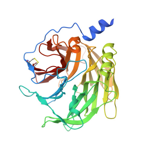
Catalytic versatility and backups in enzyme active sites: the case of serum paraoxonase 1.

Ben-David, M.Elias, M.Filippi, J.J.Dunach, E.Silman, I.Sussman, J.L.Tawfik, D.S.

(2012) J Mol Biol 418: 181-196

  • DOI: https://doi.org/10.1016/j.jmb.2012.02.042
  • Primary Citation of Related Structures:  
    3SRE, 3SRG

  • PubMed Abstract: 

    The origins of enzyme specificity are well established. However, the molecular details underlying the ability of a single active site to promiscuously bind different substrates and catalyze different reactions remain largely unknown. To better understand the molecular basis of enzyme promiscuity, we studied the mammalian serum paraoxonase 1 (PON1) whose native substrates are lipophilic lactones. We describe the crystal structures of PON1 at a catalytically relevant pH and of its complex with a lactone analogue. The various PON1 structures and the analysis of active-site mutants guided the generation of docking models of the various substrates and their reaction intermediates. The models suggest that promiscuity is driven by coincidental overlaps between the reactive intermediate for the native lactonase reaction and the ground and/or intermediate states of the promiscuous reactions. This overlap is also enabled by different active-site conformations: the lactonase activity utilizes one active-site conformation whereas the promiscuous phosphotriesterase activity utilizes another. The hydrolysis of phosphotriesters, and of the aromatic lactone dihydrocoumarin, is also driven by an alternative catalytic mode that uses only a subset of the active-site residues utilized for lactone hydrolysis. Indeed, PON1's active site shows a remarkable level of networking and versatility whereby multiple residues share the same task and individual active-site residues perform multiple tasks (e.g., binding the catalytic calcium and activating the hydrolytic water). Overall, the coexistence of multiple conformations and alternative catalytic modes within the same active site underlines PON1's promiscuity and evolutionary potential.


  • Organizational Affiliation

    Department of Structural Biology, Weizmann Institute of Science, Rehovot 76100, Israel.


Macromolecules
Find similar proteins by:  (by identity cutoff)  |  3D Structure
Entity ID: 1
MoleculeChains Sequence LengthOrganismDetailsImage
serum paraoxonase355synthetic constructMutation(s): 0 
EC: 3.1.1.2
Entity Groups  
Sequence Clusters30% Identity50% Identity70% Identity90% Identity95% Identity100% Identity
Sequence Annotations
Expand
  • Reference Sequence
Small Molecules
Ligands 5 Unique
IDChains Name / Formula / InChI Key2D Diagram3D Interactions
LMT
Query on LMT

Download Ideal Coordinates CCD File 
E [auth A]DODECYL-BETA-D-MALTOSIDE
C24 H46 O11
NLEBIOOXCVAHBD-QKMCSOCLSA-N
OCH
Query on OCH

Download Ideal Coordinates CCD File 
D [auth A]QUINOLIN-2(1H)-ONE
C9 H7 N O
LISFMEBWQUVKPJ-UHFFFAOYSA-N
BR
Query on BR

Download Ideal Coordinates CCD File 
F [auth A]BROMIDE ION
Br
CPELXLSAUQHCOX-UHFFFAOYSA-M
CA
Query on CA

Download Ideal Coordinates CCD File 
B [auth A],
C [auth A],
I [auth A]
CALCIUM ION
Ca
BHPQYMZQTOCNFJ-UHFFFAOYSA-N
CL
Query on CL

Download Ideal Coordinates CCD File 
G [auth A],
H [auth A]
CHLORIDE ION
Cl
VEXZGXHMUGYJMC-UHFFFAOYSA-M
Binding Affinity Annotations 
IDSourceBinding Affinity
OCH PDBBind:  3SRG Ki: 2000 (nM) from 1 assay(s)
Experimental Data & Validation

Experimental Data

  • Method: X-RAY DIFFRACTION
  • Resolution: 2.19 Å
  • R-Value Free: 0.227 
  • R-Value Work: 0.182 
  • R-Value Observed: 0.184 
  • Space Group: P 43 21 2
Unit Cell:
Length ( Å )Angle ( ˚ )
a = 93.544α = 90
b = 93.544β = 90
c = 144.621γ = 90
Software Package:
Software NamePurpose
HKL-2000data collection
PHASERphasing
REFMACrefinement
HKL-2000data reduction
HKL-2000data scaling

Structure Validation

View Full Validation Report


Ligand Structure Quality Assessment 


Entry History 

Deposition Data

Revision History  (Full details and data files)

  • Version 1.0: 2012-03-21
    Type: Initial release
  • Version 1.1: 2012-04-18
    Changes: Database references
  • Version 1.2: 2023-09-13
    Changes: Data collection, Database references, Derived calculations, Refinement description