3SE6

Crystal structure of the human Endoplasmic Reticulum Aminopeptidase 2


Experimental Data Snapshot

  • Method: X-RAY DIFFRACTION
  • Resolution: 3.08 Å
  • R-Value Free: 0.277 
  • R-Value Work: 0.212 
  • R-Value Observed: 0.215 

wwPDB Validation   3D Report Full Report


Ligand Structure Quality Assessment 


This is version 2.0 of the entry. See complete history


Literature

The crystal structure of human endoplasmic reticulum aminopeptidase 2 reveals the atomic basis for distinct roles in antigen processing.

Birtley, J.R.Saridakis, E.Stratikos, E.Mavridis, I.M.

(2012) Biochemistry 51: 286-295

  • DOI: https://doi.org/10.1021/bi201230p
  • Primary Citation of Related Structures:  
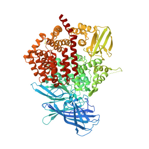
    3SE6

  • PubMed Abstract: 

    Endoplasmic reticulum aminopeptidases ERAP1 and ERAP2 cooperate to trim a vast variety of antigenic peptide precursors to generate mature epitopes for binding to major histocompatibility class I molecules. We report here the first structure of ERAP2 determined at 3.08 Å by X-ray crystallography. On the basis of residual electron density, a lysine residue has been modeled in the active site of the enzyme; thus, the structure corresponds to an enzyme-product complex. The overall domain organization is highly similar to that of the recently determined structure of ERAP1 in its closed conformation. A large internal cavity adjacent to the catalytic site can accommodate large peptide substrates. The ERAP2 structure provides a structural explanation for the different peptide N-terminal specificities between ERAP1 and ERAP2 and suggests that such differences extend throughout the whole peptide sequence. A noncrystallographic dimer observed may constitute a model for a proposed ERAP1-ERAP2 heterodimer. Overall, the structure helps explain how two homologous aminopeptidases cooperate to process a large variety of sequences, a key property of their biological role.


  • Organizational Affiliation

    Structural and Supramolecular Chemistry Laboratory, Institute of Physical Chemistry, National Center for Scientific Research Demokritos, Aghia Paraskevi 15310, Athens, Greece.


Macromolecules
Find similar proteins by:  (by identity cutoff)  |  3D Structure
Entity ID: 1
MoleculeChains Sequence LengthOrganismDetailsImage
Endoplasmic reticulum aminopeptidase 2
A, B
967Homo sapiensMutation(s): 1 
Gene Names: ERAP2LRAP
EC: 3.4.11
Membrane Entity: Yes 
UniProt & NIH Common Fund Data Resources
Find proteins for Q6P179 (Homo sapiens)
Explore Q6P179 
Go to UniProtKB:  Q6P179
PHAROS:  Q6P179
GTEx:  ENSG00000164308 
Entity Groups  
Sequence Clusters30% Identity50% Identity70% Identity90% Identity95% Identity100% Identity
UniProt GroupQ6P179
Sequence Annotations
Expand
  • Reference Sequence
Oligosaccharides

Help

Entity ID: 2
MoleculeChains Length2D Diagram Glycosylation3D Interactions
2-acetamido-2-deoxy-beta-D-glucopyranose-(1-4)-2-acetamido-2-deoxy-beta-D-glucopyranose
C, D
2N-Glycosylation
Glycosylation Resources
GlyTouCan:  G42666HT
GlyCosmos:  G42666HT
GlyGen:  G42666HT
Entity ID: 3
MoleculeChains Length2D Diagram Glycosylation3D Interactions
alpha-D-mannopyranose-(1-4)-alpha-D-mannopyranose-(1-4)-2-acetamido-2-deoxy-beta-D-glucopyranose-(1-4)-2-acetamido-2-deoxy-beta-D-glucopyranose
E
4N-Glycosylation
Glycosylation Resources
GlyTouCan:  G81874LD
GlyCosmos:  G81874LD
GlyGen:  G81874LD
Small Molecules
Ligands 4 Unique
IDChains Name / Formula / InChI Key2D Diagram3D Interactions
NAG
Query on NAG

Download Ideal Coordinates CCD File 
H [auth A],
I [auth A],
N [auth B]
2-acetamido-2-deoxy-beta-D-glucopyranose
C8 H15 N O6
OVRNDRQMDRJTHS-FMDGEEDCSA-N
MES
Query on MES

Download Ideal Coordinates CCD File 
J [auth A],
M [auth B]
2-(N-MORPHOLINO)-ETHANESULFONIC ACID
C6 H13 N O4 S
SXGZJKUKBWWHRA-UHFFFAOYSA-N
LYS
Query on LYS

Download Ideal Coordinates CCD File 
G [auth A],
K [auth B]
LYSINE
C6 H15 N2 O2
KDXKERNSBIXSRK-YFKPBYRVSA-O
ZN
Query on ZN

Download Ideal Coordinates CCD File 
F [auth A],
L [auth B]
ZINC ION
Zn
PTFCDOFLOPIGGS-UHFFFAOYSA-N
Experimental Data & Validation

Experimental Data

  • Method: X-RAY DIFFRACTION
  • Resolution: 3.08 Å
  • R-Value Free: 0.277 
  • R-Value Work: 0.212 
  • R-Value Observed: 0.215 
  • Space Group: P 1 21 1
Unit Cell:
Length ( Å )Angle ( ˚ )
a = 74.579α = 90
b = 134.362β = 90.71
c = 128.009γ = 90
Software Package:
Software NamePurpose
DNAdata collection
MOLREPphasing
PHENIXrefinement
DENZOdata reduction
SCALEPACKdata scaling

Structure Validation

View Full Validation Report


Ligand Structure Quality Assessment 


Entry History 

Deposition Data

Revision History  (Full details and data files)

  • Version 1.0: 2011-12-21
    Type: Initial release
  • Version 1.1: 2012-03-21
    Changes: Database references
  • Version 1.2: 2018-01-24
    Changes: Advisory, Structure summary
  • Version 2.0: 2020-07-29
    Type: Remediation
    Reason: Carbohydrate remediation
    Changes: Advisory, Atomic model, Data collection, Database references, Derived calculations, Structure summary