3QOQ

Crystal Structure of the Transcription Factor AmrZ in Complex with the 18 Base Pair amrZ1 Binding Site


Experimental Data Snapshot

  • Method: X-RAY DIFFRACTION
  • Resolution: 3.10 Å
  • R-Value Free: 0.295 
  • R-Value Work: 0.261 
  • R-Value Observed: 0.263 

wwPDB Validation   3D Report Full Report


This is version 1.3 of the entry. See complete history


Literature

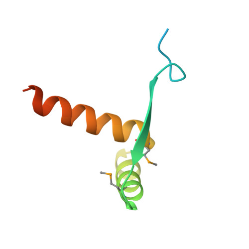
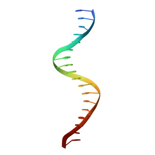
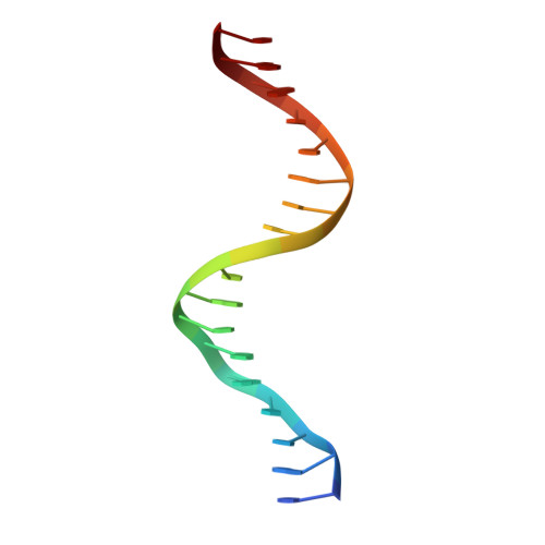
The Transcription Factor AmrZ Utilizes Multiple DNA Binding Modes to Recognize Activator and Repressor Sequences of Pseudomonas aeruginosa Virulence Genes.

Pryor Jr., E.E.Waligora, E.A.Xu, B.Dellos-Nolan, S.Wozniak, D.J.Hollis, T.

(2012) PLoS Pathog 8: e1002648-e1002648

  • DOI: https://doi.org/10.1371/journal.ppat.1002648
  • Primary Citation of Related Structures:  
    3QOQ

  • PubMed Abstract: 

    AmrZ, a member of the Ribbon-Helix-Helix family of DNA binding proteins, functions as both a transcriptional activator and repressor of multiple genes encoding Pseudomonas aeruginosa virulence factors. The expression of these virulence factors leads to chronic and sustained infections associated with worsening prognosis. In this study, we present the X-ray crystal structure of AmrZ in complex with DNA containing the repressor site, amrZ1. Binding of AmrZ to this site leads to auto-repression. AmrZ binds this DNA sequence as a dimer-of-dimers, and makes specific base contacts to two half sites, separated by a five base pair linker region. Analysis of the linker region shows a narrowing of the minor groove, causing significant distortions. AmrZ binding assays utilizing sequences containing variations in this linker region reveals that secondary structure of the DNA, conferred by the sequence of this region, is an important determinant in binding affinity. The results from these experiments allow for the creation of a model where both intrinsic structure of the DNA and specific nucleotide recognition are absolutely necessary for binding of the protein. We also examined AmrZ binding to the algD promoter, which results in activation of the alginate exopolysaccharide biosynthetic operon, and found the protein utilizes different interactions with this site. Finally, we tested the in vivo effects of this differential binding by switching the AmrZ binding site at algD, where it acts as an activator, for a repressor binding sequence and show that differences in binding alone do not affect transcriptional regulation.


  • Organizational Affiliation

    Department of Biochemistry and Center for Structural Biology, Wake Forest School of Medicine, Winston-Salem, North Carolina, United States of America.


Macromolecules

Find similar proteins by:  (by identity cutoff)  |  3D Structure
Entity ID: 1
MoleculeChains Sequence LengthOrganismDetailsImage
Alginate and motility regulator Z
A, B, C, D
69Pseudomonas aeruginosaMutation(s): 0 
Gene Names: algZamrZPA3385
UniProt
Find proteins for G3XCY4 (Pseudomonas aeruginosa (strain ATCC 15692 / DSM 22644 / CIP 104116 / JCM 14847 / LMG 12228 / 1C / PRS 101 / PAO1))
Explore G3XCY4 
Go to UniProtKB:  G3XCY4
Entity Groups  
Sequence Clusters30% Identity50% Identity70% Identity90% Identity95% Identity100% Identity
UniProt GroupG3XCY4
Sequence Annotations
Expand
  • Reference Sequence

Find similar nucleic acids by:  Sequence   |   3D Structure  

Entity ID: 2
MoleculeChains LengthOrganismImage
DNA (5'-D(*AP*CP*TP*GP*GP*CP*AP*AP*AP*AP*CP*GP*CP*CP*GP*GP*CP*A)-3')18N/A
Sequence Annotations
Expand
  • Reference Sequence

Find similar nucleic acids by:  Sequence   |   3D Structure  

Entity ID: 3
MoleculeChains LengthOrganismImage
DNA (5'-D(*TP*GP*CP*CP*GP*GP*CP*GP*TP*TP*TP*TP*GP*CP*CP*AP*GP*T)-3')18N/A
Sequence Annotations
Expand
  • Reference Sequence
Small Molecules
Modified Residues  1 Unique
IDChains TypeFormula2D DiagramParent
MSE
Query on MSE
A, B, C, D
L-PEPTIDE LINKINGC5 H11 N O2 SeMET
Experimental Data & Validation

Experimental Data

  • Method: X-RAY DIFFRACTION
  • Resolution: 3.10 Å
  • R-Value Free: 0.295 
  • R-Value Work: 0.261 
  • R-Value Observed: 0.263 
  • Space Group: I 4 2 2
Unit Cell:
Length ( Å )Angle ( ˚ )
a = 129.465α = 90
b = 129.465β = 90
c = 152.554γ = 90
Software Package:
Software NamePurpose
DENZOdata reduction
SCALEPACKdata scaling
SOLVEphasing
RESOLVEphasing
REFMACrefinement
PDB_EXTRACTdata extraction
HKL-2000data scaling

Structure Validation

View Full Validation Report


Entry History 

Deposition Data

Revision History  (Full details and data files)

  • Version 1.0: 2012-02-15
    Type: Initial release
  • Version 1.1: 2012-05-02
    Changes: Database references
  • Version 1.2: 2012-10-17
    Changes: Structure summary
  • Version 1.3: 2017-11-08
    Changes: Refinement description