


3QLU
Crystal structure of the GluK2/GluK5 (GluR6/KA2) ATD dimer assembly
- PDB DOI: https://doi.org/10.2210/pdb3QLU/pdb
- Classification: MEMBRANE PROTEIN
- Organism(s): Rattus norvegicus
- Expression System: Homo sapiens
- Mutation(s): Yes
- Deposited: 2011-02-03 Released: 2011-08-03
Experimental Data Snapshot
- Method: X-RAY DIFFRACTION
- Resolution: 2.91 Å
- R-Value Free: 0.256
- R-Value Work: 0.197
- R-Value Observed: 0.200

This is version 1.4 of the entry. See complete history.

Macromolecules
Find similar proteins by:
(by identity cutoff) | 3D Structure
Entity ID: 1 | |||||
|---|---|---|---|---|---|
| Molecule | Chains | Sequence Length | Organism | Details | Image |
| Glutamate receptor, ionotropic kainate 5 | 393 | Rattus norvegicus | Mutation(s): 0 Gene Names: Grik5 | 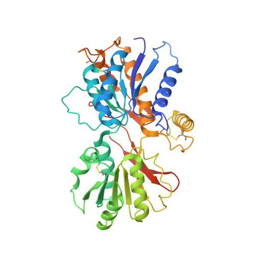 | |
UniProt | |||||
Find proteins for Q63273 (Rattus norvegicus) Explore Q63273 Go to UniProtKB: Q63273 | |||||
Entity Groups | |||||
| Sequence Clusters | 30% Identity50% Identity70% Identity90% Identity95% Identity100% Identity | ||||
| UniProt Group | Q63273 | ||||
Sequence AnnotationsExpand | |||||
| |||||
Find similar proteins by:
(by identity cutoff) | 3D Structure
Entity ID: 2 | |||||
|---|---|---|---|---|---|
| Molecule | Chains | Sequence Length | Organism | Details | Image |
| Glutamate receptor, ionotropic kainate 2 | 395 | Rattus norvegicus | Mutation(s): 2 Gene Names: Glur6, Grik2 |  | |
UniProt | |||||
Find proteins for P42260 (Rattus norvegicus) Explore P42260 Go to UniProtKB: P42260 | |||||
Entity Groups | |||||
| Sequence Clusters | 30% Identity50% Identity70% Identity90% Identity95% Identity100% Identity | ||||
| UniProt Group | P42260 | ||||
Sequence AnnotationsExpand | |||||
| |||||
Small Molecules
| Ligands 3 Unique | |||||
|---|---|---|---|---|---|
| ID | Chains | Name / Formula / InChI Key | 2D Diagram | 3D Interactions | |
| NAG Query on NAG | E [auth A] F [auth A] G [auth A] H [auth A] I [auth A] | 2-acetamido-2-deoxy-beta-D-glucopyranose C8 H15 N O6 OVRNDRQMDRJTHS-FMDGEEDCSA-N |  | ||
| GOL Query on GOL | P [auth B] | GLYCEROL C3 H8 O3 PEDCQBHIVMGVHV-UHFFFAOYSA-N |  | ||
| CL Query on CL | O [auth B], T [auth C] | CHLORIDE ION Cl VEXZGXHMUGYJMC-UHFFFAOYSA-M |  | ||
Experimental Data & Validation
Experimental Data
- Method: X-RAY DIFFRACTION
- Resolution: 2.91 Å
- R-Value Free: 0.256
- R-Value Work: 0.197
- R-Value Observed: 0.200
- Space Group: P 21 21 21
Unit Cell:
| Length ( Å ) | Angle ( ˚ ) |
|---|---|
| a = 65.626 | α = 90 |
| b = 139.548 | β = 90 |
| c = 195.41 | γ = 90 |
| Software Name | Purpose |
|---|---|
| HKL-2000 | data collection |
| PHASER | phasing |
| PHENIX | refinement |
| HKL-2000 | data reduction |
| HKL-2000 | data scaling |
Entry History
Deposition Data
- Released Date: 2011-08-03 Deposition Author(s): Kumar, J., Mayer, M.L.
Revision History (Full details and data files)
- Version 1.0: 2011-08-03
Type: Initial release - Version 1.1: 2012-10-10
Changes: Database references - Version 1.2: 2020-07-29
Type: Remediation
Reason: Carbohydrate remediation
Changes: Advisory, Data collection, Database references, Derived calculations, Structure summary - Version 1.3: 2021-03-31
Changes: Source and taxonomy, Structure summary - Version 1.4: 2023-09-13
Changes: Advisory, Data collection, Database references, Refinement description
