3QHZ

Crystal Structure of human anti-influenza Fab 2D1


Experimental Data Snapshot

  • Method: X-RAY DIFFRACTION
  • Resolution: 1.55 Å
  • R-Value Free: 0.206 
  • R-Value Work: 0.180 
  • R-Value Observed: 0.181 

wwPDB Validation   3D Report Full Report


This is version 2.1 of the entry. See complete history


Literature

An insertion mutation that distorts antibody binding site architecture enhances function of a human antibody.

Krause, J.C.Ekiert, D.C.Tumpey, T.M.Smith, P.B.Wilson, I.A.Crowe, J.E.

(2011) mBio 2: e00345-e00310

  • DOI: https://doi.org/10.1128/mBio.00345-10
  • Primary Citation of Related Structures:  
    3QHF, 3QHZ

  • PubMed Abstract: 

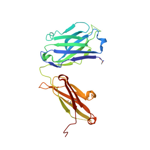
    The structural and functional significance of somatic insertions and deletions in antibody chains is unclear. Here, we demonstrate that a naturally occurring three-amino-acid insertion within the influenza virus-specific human monoclonal antibody 2D1 heavy-chain variable region reconfigures the antibody-combining site and contributes to its high potency against the 1918 and 2009 pandemic H1N1 influenza viruses. The insertion arose through a series of events, including a somatic point mutation in a predicted hot-spot motif, introduction of a new hot-spot motif, a molecular duplication due to polymerase slippage, a deletion due to misalignment, and additional somatic point mutations. Atomic resolution structures of the wild-type antibody and a variant in which the insertion was removed revealed that the three-amino-acid insertion near the base of heavy-chain complementarity-determining region (CDR) H2 resulted in a bulge in that loop. This enlarged CDR H2 loop impinges on adjacent regions, causing distortion of the CDR H1 architecture and its displacement away from the antigen-combining site. Removal of the insertion restores the canonical structure of CDR H1 and CDR H2, but binding, neutralization activity, and in vivo activity were reduced markedly because of steric conflict of CDR H1 with the hemagglutinin antigen.


  • Organizational Affiliation

    Department of Pediatrics, Vanderbilt University Medical Center, Nashville, Tennessee, USA.


Macromolecules
Find similar proteins by:  (by identity cutoff)  |  3D Structure
Entity ID: 1
MoleculeChains Sequence LengthOrganismDetailsImage
Human monoclonal antibody del2D1, Fab Heavy ChainA [auth H],
C [auth I]
232Homo sapiensMutation(s): 0 
Gene Names: Immunoglobulin gamma
Entity Groups  
Sequence Clusters30% Identity50% Identity70% Identity90% Identity95% Identity100% Identity
Sequence Annotations
Expand
  • Reference Sequence
Find similar proteins by:  (by identity cutoff)  |  3D Structure
Entity ID: 2
MoleculeChains Sequence LengthOrganismDetailsImage
Human monoclonal antibody del2D1, Fab Light ChainB [auth L],
D [auth M]
217Homo sapiensMutation(s): 0 
Gene Names: Immunoglobulin lambda
Entity Groups  
Sequence Clusters30% Identity50% Identity70% Identity90% Identity95% Identity100% Identity
Sequence Annotations
Expand
  • Reference Sequence
Small Molecules
Modified Residues  1 Unique
IDChains TypeFormula2D DiagramParent
PCA
Query on PCA
A [auth H],
C [auth I]
L-PEPTIDE LINKINGC5 H7 N O3GLN
Experimental Data & Validation

Experimental Data

  • Method: X-RAY DIFFRACTION
  • Resolution: 1.55 Å
  • R-Value Free: 0.206 
  • R-Value Work: 0.180 
  • R-Value Observed: 0.181 
  • Space Group: P 1 21 1
Unit Cell:
Length ( Å )Angle ( ˚ )
a = 69.92α = 90
b = 80.94β = 100.14
c = 77.195γ = 90
Software Package:
Software NamePurpose
Blu-Icedata collection
PHASERphasing
PHENIXrefinement
HKL-2000data reduction
HKL-2000data scaling

Structure Validation

View Full Validation Report


Entry History 

Deposition Data

Revision History  (Full details and data files)

  • Version 1.0: 2011-02-16
    Type: Initial release
  • Version 1.1: 2011-07-13
    Changes: Version format compliance
  • Version 2.0: 2019-12-25
    Changes: Derived calculations, Polymer sequence
  • Version 2.1: 2023-09-13
    Changes: Data collection, Database references, Refinement description