3QFC

Crystal Structure of Human NADPH-Cytochrome P450 (V492E mutant)


Experimental Data Snapshot

  • Method: X-RAY DIFFRACTION
  • Resolution: 1.80 Å
  • R-Value Free: 0.265 
  • R-Value Work: 0.232 
  • R-Value Observed: 0.232 

wwPDB Validation   3D Report Full Report


Ligand Structure Quality Assessment 


This is version 1.3 of the entry. See complete history


Literature

Structural basis for human NADPH-cytochrome P450 oxidoreductase deficiency.

Xia, C.Panda, S.P.Marohnic, C.C.Martasek, P.Masters, B.S.Kim, J.J.

(2011) Proc Natl Acad Sci U S A 108: 13486-13491

  • DOI: https://doi.org/10.1073/pnas.1106632108
  • Primary Citation of Related Structures:  
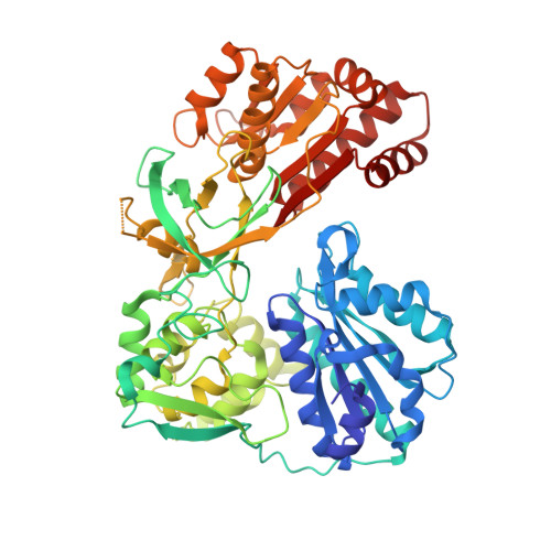
    3QE2, 3QFC, 3QFR, 3QFS, 3QFT

  • PubMed Abstract: 

    NADPH-cytochrome P450 oxidoreductase (CYPOR) is essential for electron donation to microsomal cytochrome P450-mediated monooxygenation in such diverse physiological processes as drug metabolism (approximately 85-90% of therapeutic drugs), steroid biosynthesis, and bioactive metabolite production (vitamin D and retinoic acid metabolites). Expressed by a single gene, CYPOR's role with these multiple redox partners renders it a model for understanding protein-protein interactions at the structural level. Polymorphisms in human CYPOR have been shown to lead to defects in bone development and steroidogenesis, resulting in sexual dimorphisms, the severity of which differs significantly depending on the degree of CYPOR impairment. The atomic structure of human CYPOR is presented, with structures of two naturally occurring missense mutations, V492E and R457H. The overall structures of these CYPOR variants are similar to wild type. However, in both variants, local disruption of H bonding and salt bridging, involving the FAD pyrophosphate moiety, leads to weaker FAD binding, unstable protein, and loss of catalytic activity, which can be rescued by cofactor addition. The modes of polypeptide unfolding in these two variants differ significantly, as revealed by limited trypsin digestion: V492E is less stable but unfolds locally and gradually, whereas R457H is more stable but unfolds globally. FAD addition to either variant prevents trypsin digestion, supporting the role of the cofactor in conferring stability to CYPOR structure. Thus, CYPOR dysfunction in patients harboring these particular mutations may possibly be prevented by riboflavin therapy in utero, if predicted prenatally, or rescued postnatally in less severe cases.


  • Organizational Affiliation

    Department of Biochemistry, Medical College of Wisconsin, Milwaukee, WI 53226, USA.


Macromolecules
Find similar proteins by:  (by identity cutoff)  |  3D Structure
Entity ID: 1
MoleculeChains Sequence LengthOrganismDetailsImage
NADPH--cytochrome P450 reductase
A, B
618Homo sapiensMutation(s): 1 
Gene Names: PORCYPOR
EC: 1.6.2.4
UniProt & NIH Common Fund Data Resources
Find proteins for P16435 (Homo sapiens)
Explore P16435 
Go to UniProtKB:  P16435
PHAROS:  P16435
GTEx:  ENSG00000127948 
Entity Groups  
Sequence Clusters30% Identity50% Identity70% Identity90% Identity95% Identity100% Identity
UniProt GroupP16435
Sequence Annotations
Expand
  • Reference Sequence
Small Molecules
Ligands 4 Unique
IDChains Name / Formula / InChI Key2D Diagram3D Interactions
FAD
Query on FAD

Download Ideal Coordinates CCD File 
C [auth A],
G [auth B]
FLAVIN-ADENINE DINUCLEOTIDE
C27 H33 N9 O15 P2
VWWQXMAJTJZDQX-UYBVJOGSSA-N
NAP
Query on NAP

Download Ideal Coordinates CCD File 
E [auth A],
I [auth B]
NADP NICOTINAMIDE-ADENINE-DINUCLEOTIDE PHOSPHATE
C21 H28 N7 O17 P3
XJLXINKUBYWONI-NNYOXOHSSA-N
FMN
Query on FMN

Download Ideal Coordinates CCD File 
D [auth A],
H [auth B]
FLAVIN MONONUCLEOTIDE
C17 H21 N4 O9 P
FVTCRASFADXXNN-SCRDCRAPSA-N
CA
Query on CA

Download Ideal Coordinates CCD File 
F [auth A]CALCIUM ION
Ca
BHPQYMZQTOCNFJ-UHFFFAOYSA-N
Experimental Data & Validation

Experimental Data

  • Method: X-RAY DIFFRACTION
  • Resolution: 1.80 Å
  • R-Value Free: 0.265 
  • R-Value Work: 0.232 
  • R-Value Observed: 0.232 
  • Space Group: P 21 21 21
Unit Cell:
Length ( Å )Angle ( ˚ )
a = 70.716α = 90
b = 118.508β = 90
c = 155.36γ = 90
Software Package:
Software NamePurpose
HKL-2000data collection
MOLREPphasing
CNSrefinement
HKL-2000data reduction
HKL-2000data scaling

Structure Validation

View Full Validation Report


Ligand Structure Quality Assessment 


Entry History 

Deposition Data

Revision History  (Full details and data files)

  • Version 1.0: 2011-08-03
    Type: Initial release
  • Version 1.1: 2011-08-24
    Changes: Database references
  • Version 1.2: 2011-08-31
    Changes: Database references
  • Version 1.3: 2023-09-13
    Changes: Data collection, Database references, Derived calculations, Refinement description