3PXU

Crystal structure of phosphopantetheine adenylyltransferase from Burkholderia pseudomallei bound to dephospho-coenzyme A


Experimental Data Snapshot

  • Method: X-RAY DIFFRACTION
  • Resolution: 2.10 Å
  • R-Value Free: 0.242 
  • R-Value Work: 0.200 
  • R-Value Observed: 0.202 

wwPDB Validation   3D Report Full Report


Ligand Structure Quality Assessment 


This is version 1.5 of the entry. See complete history


Literature

Structures of phosphopantetheine adenylyltransferase from Burkholderia pseudomallei.

Edwards, T.E.Leibly, D.J.Bhandari, J.Statnekov, J.B.Phan, I.Dieterich, S.H.Abendroth, J.Staker, B.L.Van Voorhis, W.C.Myler, P.J.Stewart, L.J.

(2011) Acta Crystallogr Sect F Struct Biol Cryst Commun 67: 1032-1037

  • DOI: https://doi.org/10.1107/S1744309111004349
  • Primary Citation of Related Structures:  
    3K9W, 3PXU

  • PubMed Abstract: 

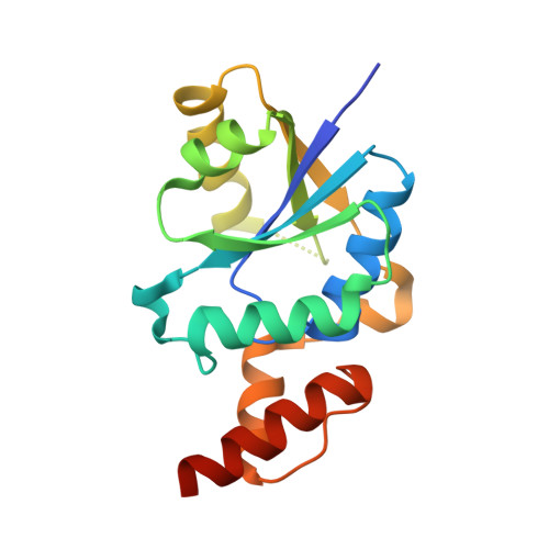
    Phosphopantetheine adenylyltransferase (PPAT) catalyzes the fourth of five steps in the coenzyme A biosynthetic pathway, reversibly transferring an adenylyl group from ATP onto 4'-phosphopantetheine to yield dephospho-coenzyme A and pyrophosphate. Burkholderia pseudomallei is a soil- and water-borne pathogenic bacterium and the etiologic agent of melioidosis, a potentially fatal systemic disease present in southeast Asia. Two crystal structures are presented of the PPAT from B. pseudomallei with the expectation that, because of the importance of the enzyme in coenzyme A biosynthesis, they will aid in the search for defenses against this pathogen. A crystal grown in ammonium sulfate yielded a 2.1 Å resolution structure that contained dephospho-coenzyme A with partial occupancy. The overall structure and ligand-binding interactions are quite similar to other bacterial PPAT crystal structures. A crystal grown at low pH in the presence of coenzyme A yielded a 1.6 Å resolution structure in the same crystal form. However, the experimental electron density was not reflective of fully ordered coenzyme A, but rather was only reflective of an ordered 4'-diphosphopantetheine moiety.


  • Organizational Affiliation

    Seattle Structural Genomics Center for Infectious Disease (SSGCID), USA. tedwards@embios.com


Macromolecules
Find similar proteins by:  (by identity cutoff)  |  3D Structure
Entity ID: 1
MoleculeChains Sequence LengthOrganismDetailsImage
Phosphopantetheine adenylyltransferase170Burkholderia pseudomallei 1710bMutation(s): 0 
Gene Names: BURPS1710b_0748coaDcoaD BURPS1710B_0748
EC: 2.7.7.3
UniProt
Find proteins for Q3JW91 (Burkholderia pseudomallei (strain 1710b))
Explore Q3JW91 
Go to UniProtKB:  Q3JW91
Entity Groups  
Sequence Clusters30% Identity50% Identity70% Identity90% Identity95% Identity100% Identity
UniProt GroupQ3JW91
Sequence Annotations
Expand
  • Reference Sequence
Experimental Data & Validation

Experimental Data

  • Method: X-RAY DIFFRACTION
  • Resolution: 2.10 Å
  • R-Value Free: 0.242 
  • R-Value Work: 0.200 
  • R-Value Observed: 0.202 
  • Space Group: I 4 3 2
Unit Cell:
Length ( Å )Angle ( ˚ )
a = 134.161α = 90
b = 134.161β = 90
c = 134.161γ = 90
Software Package:
Software NamePurpose
DENZOdata reduction
SCALEPACKdata scaling
PHASERphasing
REFMACrefinement
PDB_EXTRACTdata extraction
StructureStudiodata collection
HKL-2000data reduction
HKL-2000data scaling

Structure Validation

View Full Validation Report


Ligand Structure Quality Assessment 


Entry History 

Deposition Data

Revision History  (Full details and data files)

  • Version 1.0: 2010-12-22
    Type: Initial release
  • Version 1.1: 2011-07-13
    Changes: Version format compliance
  • Version 1.2: 2011-09-21
    Changes: Database references
  • Version 1.3: 2013-10-30
    Changes: Database references
  • Version 1.4: 2017-11-08
    Changes: Refinement description
  • Version 1.5: 2023-09-13
    Changes: Data collection, Database references, Derived calculations, Refinement description