3PS2

Crystal structure of the Escherichia Coli LPXC/LPC-012 complex


Experimental Data Snapshot

  • Method: X-RAY DIFFRACTION
  • Resolution: 2.30 Å
  • R-Value Free: 0.261 
  • R-Value Work: 0.207 
  • R-Value Observed: 0.210 

wwPDB Validation   3D Report Full Report


Ligand Structure Quality Assessment 


This is version 1.4 of the entry. See complete history


Literature

Syntheses, structures and antibiotic activities of LpxC inhibitors based on the diacetylene scaffold.

Liang, X.Lee, C.J.Chen, X.Chung, H.S.Zeng, D.Raetz, C.R.Li, Y.Zhou, P.Toone, E.J.

(2011) Bioorg Med Chem 19: 852-860

  • DOI: https://doi.org/10.1016/j.bmc.2010.12.017
  • Primary Citation of Related Structures:  
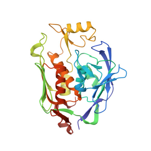
    3PS1, 3PS2, 3PS3

  • PubMed Abstract: 

    Compounds inhibiting LpxC in the lipid A biosynthetic pathway are promising leads for novel antibiotics against multidrug-resistant Gram-negative pathogens. We report the syntheses and structural and biochemical characterizations of LpxC inhibitors based on a diphenyl-diacetylene (1,4-diphenyl-1,3-butadiyne) threonyl-hydroxamate scaffold. These studies provide a molecular interpretation for the differential antibiotic activities of compounds with a substituted distal phenyl ring as well as the absolute stereochemical requirement at the C2, but not C3, position of the threonyl group.


  • Organizational Affiliation

    Department of Chemistry, Jilin University, Changchun, PR China.


Macromolecules
Find similar proteins by:  (by identity cutoff)  |  3D Structure
Entity ID: 1
MoleculeChains Sequence LengthOrganismDetailsImage
UDP-3-O-[3-hydroxymyristoyl] N-acetylglucosamine deacetylase300Escherichia coli IHE3034Mutation(s): 0 
Gene Names: ECOK1_0097ENVAlpxC
EC: 3.5.1
Entity Groups  
Sequence Clusters30% Identity50% Identity70% Identity90% Identity95% Identity100% Identity
Sequence Annotations
Expand
  • Reference Sequence
Small Molecules
Ligands 5 Unique
IDChains Name / Formula / InChI Key2D Diagram3D Interactions
ZH4
Query on ZH4

Download Ideal Coordinates CCD File 
C [auth A]4-[4-(3-aminophenyl)buta-1,3-diyn-1-yl]-N-[(2S,3R)-3-hydroxy-1-nitroso-1-oxobutan-2-yl]benzamide
C21 H17 N3 O4
HOPLGMFTTVLMDZ-KUHUBIRLSA-N
UKW
Query on UKW

Download Ideal Coordinates CCD File 
D [auth A]4-ethynyl-N-[(1S,2R)-2-hydroxy-1-(oxocarbamoyl)propyl]benzamide
C13 H12 N2 O4
JJXZDQZGPLQGCP-KCJUWKMLSA-N
SO4
Query on SO4

Download Ideal Coordinates CCD File 
H [auth A],
I [auth A],
J [auth A]
SULFATE ION
O4 S
QAOWNCQODCNURD-UHFFFAOYSA-L
DMS
Query on DMS

Download Ideal Coordinates CCD File 
E [auth A],
F [auth A],
G [auth A]
DIMETHYL SULFOXIDE
C2 H6 O S
IAZDPXIOMUYVGZ-UHFFFAOYSA-N
ZN
Query on ZN

Download Ideal Coordinates CCD File 
B [auth A]ZINC ION
Zn
PTFCDOFLOPIGGS-UHFFFAOYSA-N
Experimental Data & Validation

Experimental Data

  • Method: X-RAY DIFFRACTION
  • Resolution: 2.30 Å
  • R-Value Free: 0.261 
  • R-Value Work: 0.207 
  • R-Value Observed: 0.210 
  • Space Group: P 61
Unit Cell:
Length ( Å )Angle ( ˚ )
a = 107.449α = 90
b = 107.449β = 90
c = 53.422γ = 120
Software Package:
Software NamePurpose
DENZOdata reduction
SCALEPACKdata scaling
PHASERphasing
PHENIXrefinement
PDB_EXTRACTdata extraction
HKL-2000data collection

Structure Validation

View Full Validation Report


Ligand Structure Quality Assessment 


Entry History 

Deposition Data

Revision History  (Full details and data files)

  • Version 1.0: 2011-01-19
    Type: Initial release
  • Version 1.1: 2011-07-13
    Changes: Version format compliance
  • Version 1.2: 2017-11-08
    Changes: Refinement description
  • Version 1.3: 2018-01-24
    Changes: Structure summary
  • Version 1.4: 2024-02-21
    Changes: Data collection, Database references, Derived calculations