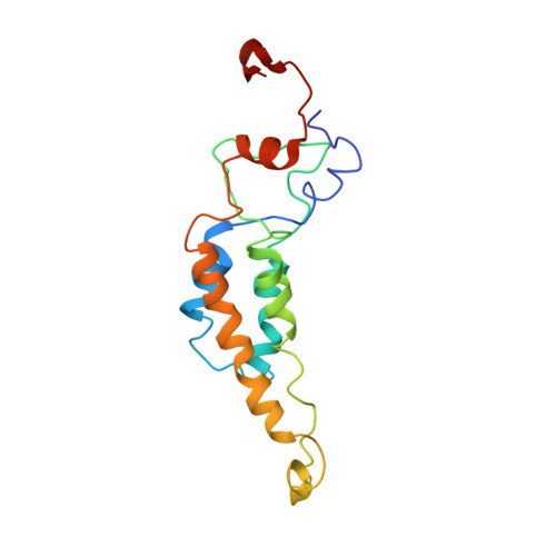
3PDM

Hibiscus Latent Singapore virus


Experimental Data Snapshot

  • Method: FIBER DIFFRACTION
  • Resolution: 3.50 Å
  • R-Value Work: 0.096 
  • R-Value Observed: 0.096 

wwPDB Validation   3D Report Full Report


This is version 1.3 of the entry. See complete history


Literature

Structure of Hibiscus latent Singapore virus by fiber diffraction: A non-conserved His122 contributes to coat protein stability

Tewary, S.K.Oda, T.Kendall, A.Bian, W.Stubbs, G.Wong, S.M.Swaminathan, K.

(2010) J Mol Biol 

  • DOI: https://doi.org/10.1016/j.jmb.2010.12.032
  • Primary Citation of Related Structures:  
    3PDM

  • PubMed Abstract: 

    Hibiscus latent Singapore virus (HLSV) is a rigid rod-shaped plant virus and a new member of the Tobamovirus family. Unlike all other Tobamoviruses, the HLSV genome contains a unique poly(A) tract in its 3' untranslated region. The virion is composed of a monomeric coat protein (CP) unit of 18 kDa, arranged as a right-handed helix around the virus axis. We have determined the structure of HLSV at 3.5 Å by X-ray fiber diffraction and refined it to an R-factor of 0.096. While the overall structure of the HLSV CP resembles that of other Tobamoviruses, there are a few unique differences. There is a kink in the LR helix due to the presence of His122. Also, the adjacent Lys123 may further destabilize the helix by positive charge repulsion, making the kink more pronounced. The His122-Asp88 salt bridge provides significant stability to the loop adjacent to the RR helix. Carboxyl-carboxylate interactions that drive viral disassembly are also different in HLSV. The nucleotide recognition mechanisms for virus assembly between HLSV and ribgrass mosaic virus are similar, but different between tobacco mosaic virus and cucumber green mottle mosaic virus.


  • Organizational Affiliation

    Department of Biological Sciences, National University of Singapore, Singapore 117543, Singapore.


Macromolecules

Find similar proteins by:  (by identity cutoff)  |  3D Structure
Entity ID: 2
MoleculeChains Sequence LengthOrganismDetailsImage
Coat proteinB [auth P]162Hibiscus latent Singapore virusMutation(s): 0 
UniProt
Find proteins for Q8BE68 (Hibiscus latent Singapore virus)
Explore Q8BE68 
Go to UniProtKB:  Q8BE68
Entity Groups  
Sequence Clusters30% Identity50% Identity70% Identity90% Identity95% Identity100% Identity
UniProt GroupQ8BE68
Sequence Annotations
Expand
  • Reference Sequence

Find similar nucleic acids by:  Sequence   |   3D Structure  

Entity ID: 1
MoleculeChains LengthOrganismImage
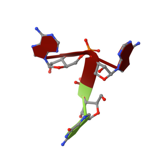
RNA (5'-R(P*GP*AP*A)-3')A [auth R]3N/A
Sequence Annotations
Expand
  • Reference Sequence
Experimental Data & Validation

Experimental Data

  • Method: FIBER DIFFRACTION
  • Resolution: 3.50 Å
  • R-Value Work: 0.096 
  • R-Value Observed: 0.096 
  • Space Group: P 1
Unit Cell:
Length ( Å )Angle ( ˚ )
a = 1α = 90
b = 1β = 90
c = 1γ = 90
Software Package:
Software NamePurpose
ADSCdata collection
X-PLORmodel building
X-PLORrefinement
WCENdata reduction
RADdata reduction
CORRdata reduction
WCENdata scaling
RADdata scaling
CORRdata scaling
X-PLORphasing

Structure Validation

View Full Validation Report


Entry History 

Deposition Data

Revision History  (Full details and data files)

  • Version 1.0: 2011-01-12
    Type: Initial release
  • Version 1.1: 2011-07-13
    Changes: Version format compliance
  • Version 1.2: 2017-11-08
    Changes: Refinement description
  • Version 1.3: 2024-03-20
    Changes: Data collection, Database references, Derived calculations