3OQ2

Structure of a CRISPR associated protein Cas2 from Desulfovibrio vulgaris


Experimental Data Snapshot

  • Method: X-RAY DIFFRACTION
  • Resolution: 1.35 Å
  • R-Value Free: 0.177 
  • R-Value Work: 0.128 
  • R-Value Observed: 0.130 

wwPDB Validation   3D Report Full Report


This is version 1.3 of the entry. See complete history


Literature

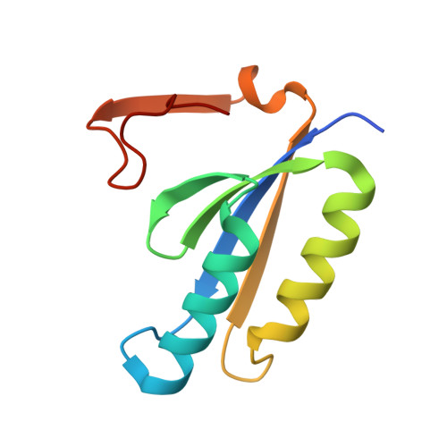
Structure of a CRISPR-associated protein Cas2 from Desulfovibrio vulgaris.

Samai, P.Smith, P.Shuman, S.

(2010) Acta Crystallogr Sect F Struct Biol Cryst Commun 66: 1552-1556

  • DOI: https://doi.org/10.1107/S1744309110039801
  • Primary Citation of Related Structures:  
    3OQ2

  • PubMed Abstract: 

    CRISPRs (clustered regularly interspaced short palindromic repeats) provide bacteria and archaea with RNA-guided acquired immunity to invasive DNAs. CRISPR-associated (Cas) proteins carry out the immune effector functions. Cas2 is a universal component of the CRISPR system. Here, a 1.35 Å resolution crystal structure of Cas2 from the bacterium Desulfovibrio vulgaris (DvuCas2) is reported. DvuCas2 is a homodimer, with each protomer consisting of an N-terminal βαββαβ ferredoxin fold (amino acids 1-78) to which is appended a C-terminal segment (amino acids 79-102) that includes a short 3(10)-helix and a fifth β-strand. The β5 strands align with the β4 strands of the opposite protomers, resulting in two five-stranded antiparallel β-sheets that form a sandwich at the dimer interface. The DvuCas2 dimer is stabilized by a distinctive network of hydrophilic cross-protomer side-chain interactions.


  • Organizational Affiliation

    Molecular Biology Program, Sloan-Kettering Institute for Cancer Research, USA.


Macromolecules
Find similar proteins by:  (by identity cutoff)  |  3D Structure
Entity ID: 1
MoleculeChains Sequence LengthOrganismDetailsImage
CRISPR-associated protein Cas2
A, B
103Nitratidesulfovibrio vulgaris str. HildenboroughMutation(s): 0 
Gene Names: cas2DVUA0135
UniProt
Find proteins for Q72WF4 (Desulfovibrio vulgaris (strain ATCC 29579 / DSM 644 / NCIMB 8303 / VKM B-1760 / Hildenborough))
Explore Q72WF4 
Go to UniProtKB:  Q72WF4
Entity Groups  
Sequence Clusters30% Identity50% Identity70% Identity90% Identity95% Identity100% Identity
UniProt GroupQ72WF4
Sequence Annotations
Expand
  • Reference Sequence
Small Molecules
Ligands 5 Unique
IDChains Name / Formula / InChI Key2D Diagram3D Interactions
CIT
Query on CIT

Download Ideal Coordinates CCD File 
I [auth A],
K [auth B],
L [auth B]
CITRIC ACID
C6 H8 O7
KRKNYBCHXYNGOX-UHFFFAOYSA-N
TRS
Query on TRS

Download Ideal Coordinates CCD File 
D [auth A]2-AMINO-2-HYDROXYMETHYL-PROPANE-1,3-DIOL
C4 H12 N O3
LENZDBCJOHFCAS-UHFFFAOYSA-O
SO4
Query on SO4

Download Ideal Coordinates CCD File 
F [auth A],
G [auth A],
H [auth A],
J [auth A],
M [auth B]
SULFATE ION
O4 S
QAOWNCQODCNURD-UHFFFAOYSA-L
CL
Query on CL

Download Ideal Coordinates CCD File 
E [auth A]CHLORIDE ION
Cl
VEXZGXHMUGYJMC-UHFFFAOYSA-M
NA
Query on NA

Download Ideal Coordinates CCD File 
C [auth A]SODIUM ION
Na
FKNQFGJONOIPTF-UHFFFAOYSA-N
Experimental Data & Validation

Experimental Data

  • Method: X-RAY DIFFRACTION
  • Resolution: 1.35 Å
  • R-Value Free: 0.177 
  • R-Value Work: 0.128 
  • R-Value Observed: 0.130 
  • Space Group: C 1 2 1
Unit Cell:
Length ( Å )Angle ( ˚ )
a = 107.874α = 90
b = 48.81β = 99.2
c = 41.32γ = 90
Software Package:
Software NamePurpose
HKL-2000data collection
SOLVEphasing
PHENIXrefinement
HKL-2000data reduction
SCALEPACKdata scaling

Structure Validation

View Full Validation Report


Entry History 

Deposition Data

Revision History  (Full details and data files)

  • Version 1.0: 2010-12-22
    Type: Initial release
  • Version 1.1: 2011-07-13
    Changes: Version format compliance
  • Version 1.2: 2017-09-13
    Changes: Refinement description
  • Version 1.3: 2024-02-21
    Changes: Data collection, Database references, Derived calculations