3OAU

Antibody 2G12 Recognizes Di-Mannose Equivalently in Domain- and Non-Domain-Exchanged Forms, but only binds the HIV-1 Glycan Shield if Domain-Exchanged


Experimental Data Snapshot

  • Method: X-RAY DIFFRACTION
  • Resolution: 1.90 Å
  • R-Value Free: 0.243 
  • R-Value Work: 0.212 
  • R-Value Observed: 0.213 

wwPDB Validation   3D Report Full Report


This is version 2.0 of the entry. See complete history


Literature

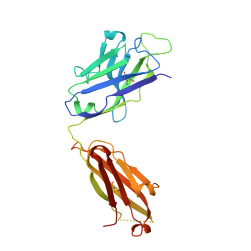
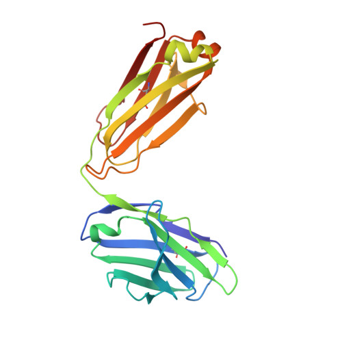
Antibody 2G12 recognizes di-mannose equivalently in domain- and nondomain-exchanged forms but only binds the HIV-1 glycan shield if domain exchanged.

Doores, K.J.Fulton, Z.Huber, M.Wilson, I.A.Burton, D.R.

(2010) J Virol 84: 10690-10699

  • DOI: https://doi.org/10.1128/JVI.01110-10
  • Primary Citation of Related Structures:  
    3OAU

  • PubMed Abstract: 

    The broadly neutralizing anti-human immunodeficiency virus type 1 (HIV-1) antibody 2G12 targets the high-mannose cluster on the glycan shield of HIV-1. 2G12 has a unique V(H) domain-exchanged structure, with a multivalent binding surface that includes two primary glycan binding sites. The high-mannose cluster is an attractive target for HIV-1 vaccine design, but so far, no carbohydrate immunogen has elicited 2G12-like antibodies. Important questions remain as to how this domain exchange arose in 2G12 and how this unusual event conferred unexpected reactivity against the glycan shield of HIV-1. In order to address these questions, we generated a nondomain-exchanged variant of 2G12 to produce a conventional Y/T-shaped antibody through a single amino acid substitution (2G12 I19R) and showed that, as for the 2G12 wild type (2G12 WT), this antibody is able to recognize the same Manα1,2Man motif on recombinant gp120, Candida albicans, and synthetic glycoconjugates. However, the nondomain-exchanged variant of 2G12 is unable to bind the cluster of mannose moieties on the surface of HIV-1. Crystallographic analysis of 2G12 I19R in complex with Manα1,2Man revealed an adaptable hinge between V(H) and C(H)1 that enables the V(H) and V(L) domains to assemble in such a way that the configuration of the primary binding site and its interaction with disaccharide are remarkably similar in the nondomain-exchanged and domain-exchanged forms. Together with data that suggest that very few substitutions are required for domain exchange, the results suggest potential mechanisms for the evolution of domain-exchanged antibodies and immunization strategies for eliciting such antibodies.


  • Organizational Affiliation

    Department of Immunology and Microbial Science, The Scripps Research Institute, 10550 North Torrey Pines Road, La Jolla, CA 92037, USA.


Macromolecules
Find similar proteins by:  (by identity cutoff)  |  3D Structure
Entity ID: 1
MoleculeChains Sequence LengthOrganismDetailsImage
Fab 2G12, heavy chainA [auth H]225Homo sapiensMutation(s): 0 
Entity Groups  
Sequence Clusters30% Identity50% Identity70% Identity90% Identity95% Identity100% Identity
Sequence Annotations
Expand
  • Reference Sequence
Find similar proteins by:  (by identity cutoff)  |  3D Structure
Entity ID: 2
MoleculeChains Sequence LengthOrganismDetailsImage
Fab 2G12, light chainB [auth L]212Homo sapiensMutation(s): 0 
Entity Groups  
Sequence Clusters30% Identity50% Identity70% Identity90% Identity95% Identity100% Identity
Sequence Annotations
Expand
  • Reference Sequence
Oligosaccharides

Help

Entity ID: 3
MoleculeChains Length2D Diagram Glycosylation3D Interactions
alpha-D-mannopyranose-(1-2)-alpha-D-mannopyranoseC [auth A]2N/A
Glycosylation Resources
GlyTouCan:  G53402KW
GlyCosmos:  G53402KW
GlyGen:  G53402KW
Biologically Interesting Molecules (External Reference) 1 Unique
Experimental Data & Validation

Experimental Data

  • Method: X-RAY DIFFRACTION
  • Resolution: 1.90 Å
  • R-Value Free: 0.243 
  • R-Value Work: 0.212 
  • R-Value Observed: 0.213 
  • Space Group: P 1
Unit Cell:
Length ( Å )Angle ( ˚ )
a = 39.301α = 92.91
b = 56.628β = 105.05
c = 57.653γ = 92.74
Software Package:
Software NamePurpose
BUSTER-TNTrefinement
PDB_EXTRACTdata extraction
BUSTERrefinement

Structure Validation

View Full Validation Report


Entry History 

Deposition Data

Revision History  (Full details and data files)

  • Version 1.0: 2011-01-12
    Type: Initial release
  • Version 1.1: 2011-07-13
    Changes: Version format compliance
  • Version 2.0: 2020-07-29
    Type: Remediation
    Reason: Carbohydrate remediation
    Changes: Atomic model, Data collection, Derived calculations, Structure summary