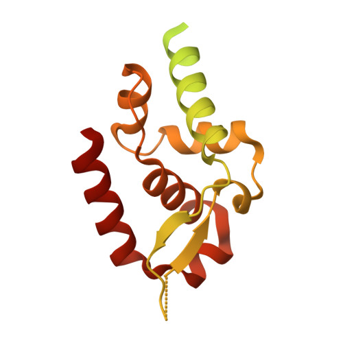
Crystal structure of phosphoprotein/Protein P/Protein M1 residues 69-297 from Rabies virus reveals degradation to C-terminal domain only
Edwards, T.E., Abendroth, J., Seattle Structural Genomics Center for Infectious Disease (SSGCID)To be published.