3O26

The structure of salutaridine reductase from Papaver somniferum.


Experimental Data Snapshot

  • Method: X-RAY DIFFRACTION
  • Resolution: 1.91 Å
  • R-Value Free: 0.208 
  • R-Value Work: 0.193 
  • R-Value Observed: 0.193 

wwPDB Validation   3D Report Full Report


Ligand Structure Quality Assessment 


This is version 1.1 of the entry. See complete history


Literature

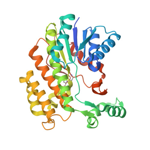
The atomic structure of salutaridine reductase from the opium poppy Papaver somniferum.

Higashi, Y.Kutchan, T.M.Smith, T.J.

(2010) J Biol Chem 66: 163-166

  • DOI: https://doi.org/10.1074/jbc.M110.168633
  • Primary Citation of Related Structures:  
    3O26

  • PubMed Abstract: 

    The opium poppy (Papaver somniferum L.) is one of the oldest known medicinal plants. In the biosynthetic pathway for morphine and codeine, salutaridine is reduced to salutaridinol by salutaridine reductase (SalR; EC 1.1.1.248) using NADPH as coenzyme. Here, we report the atomic structure of SalR to a resolution of ∼1.9 Å in the presence of NADPH. The core structure is highly homologous to other members of the short chain dehydrogenase/reductase family. The major difference is that the nicotinamide moiety and the substrate-binding pocket are covered by a loop (residues 265-279), on top of which lies a large "flap"-like domain (residues 105-140). This configuration appears to be a combination of the two common structural themes found in other members of the short chain dehydrogenase/reductase family. Previous modeling studies suggested that substrate inhibition is due to mutually exclusive productive and nonproductive modes of substrate binding in the active site. This model was tested via site-directed mutagenesis, and a number of these mutations abrogated substrate inhibition. However, the atomic structure of SalR shows that these mutated residues are instead distributed over a wide area of the enzyme, and many are not in the active site. To explain how residues distal to the active site might affect catalysis, a model is presented whereby SalR may undergo significant conformational changes during catalytic turnover.


  • Organizational Affiliation

    Donald Danforth Plant Science Center, St. Louis, Missouri 63132, USA.


Macromolecules
Find similar proteins by:  (by identity cutoff)  |  3D Structure
Entity ID: 1
MoleculeChains Sequence LengthOrganismDetailsImage
Salutaridine reductase311Papaver somniferumMutation(s): 0 
EC: 1.1.1.248
UniProt
Find proteins for Q071N0 (Papaver somniferum)
Explore Q071N0 
Go to UniProtKB:  Q071N0
Entity Groups  
Sequence Clusters30% Identity50% Identity70% Identity90% Identity95% Identity100% Identity
UniProt GroupQ071N0
Sequence Annotations
Expand
  • Reference Sequence
Small Molecules
Ligands 1 Unique
IDChains Name / Formula / InChI Key2D Diagram3D Interactions
NDP
Query on NDP

Download Ideal Coordinates CCD File 
B [auth A]NADPH DIHYDRO-NICOTINAMIDE-ADENINE-DINUCLEOTIDE PHOSPHATE
C21 H30 N7 O17 P3
ACFIXJIJDZMPPO-NNYOXOHSSA-N
Experimental Data & Validation

Experimental Data

  • Method: X-RAY DIFFRACTION
  • Resolution: 1.91 Å
  • R-Value Free: 0.208 
  • R-Value Work: 0.193 
  • R-Value Observed: 0.193 
  • Space Group: P 64 2 2
Unit Cell:
Length ( Å )Angle ( ˚ )
a = 139.906α = 90
b = 139.906β = 90
c = 100.174γ = 120
Software Package:
Software NamePurpose
DENZOdata reduction
SCALEPACKdata scaling
SOLVEphasing
PHENIXrefinement
PDB_EXTRACTdata extraction
HKL-3000data collection

Structure Validation

View Full Validation Report


Ligand Structure Quality Assessment 


Entry History 

Deposition Data

Revision History  (Full details and data files)

  • Version 1.0: 2010-12-15
    Type: Initial release
  • Version 1.1: 2011-07-13
    Changes: Version format compliance