3NZM

Crystal structure of DNAE intein with N-extein in redox trap


Experimental Data Snapshot

  • Method: X-RAY DIFFRACTION
  • Resolution: 1.55 Å
  • R-Value Free: 0.260 
  • R-Value Work: 0.220 
  • R-Value Observed: 0.220 

wwPDB Validation   3D Report Full Report


This is version 2.0 of the entry. See complete history


Literature

Structure of catalytically competent intein caught in a redox trap with functional and evolutionary implications.

Callahan, B.P.Topilina, N.I.Stanger, M.J.Van Roey, P.Belfort, M.

(2011) Nat Struct Mol Biol 18: 630-633

  • DOI: https://doi.org/10.1038/nsmb.2041
  • Primary Citation of Related Structures:  
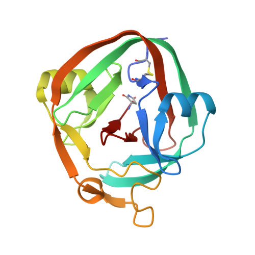
    3NZM

  • PubMed Abstract: 

    Here we describe self-splicing proteins, called inteins, that function as redox-responsive switches in bacteria. Redox regulation was achieved by engineering a disulfide bond between the intein's catalytic cysteine and a cysteine in the flanking 'extein' sequence. This interaction was validated by an X-ray structure, which includes a transient splice junction. A natural analog of the designed system was identified in Pyrococcus abyssi, suggesting an unprecedented form of adaptive, post-translational regulation.


  • Organizational Affiliation

    Wadsworth Center, New York State Department of Health, Albany, New York, USA.


Macromolecules
Find similar proteins by:  (by identity cutoff)  |  3D Structure
Entity ID: 1
MoleculeChains Sequence LengthOrganismDetailsImage
DNA polymerase III subunit alpha168Synechocystis sp. PCC 6803Mutation(s): 8 
Gene Names: dnaE-Nslr0603dnaE-Csll1572
EC: 2.7.7.7
UniProt
Find proteins for P74750 (Synechocystis sp. (strain PCC 6803 / Kazusa))
Explore P74750 
Go to UniProtKB:  P74750
Entity Groups  
Sequence Clusters30% Identity50% Identity70% Identity90% Identity95% Identity100% Identity
UniProt GroupP74750
Sequence Annotations
Expand
  • Reference Sequence
Small Molecules
Modified Residues  1 Unique
IDChains TypeFormula2D DiagramParent
SNN
Query on SNN
A
L-PEPTIDE LINKINGC4 H6 N2 O2ASN
Experimental Data & Validation

Experimental Data

  • Method: X-RAY DIFFRACTION
  • Resolution: 1.55 Å
  • R-Value Free: 0.260 
  • R-Value Work: 0.220 
  • R-Value Observed: 0.220 
  • Space Group: P 21 21 21
Unit Cell:
Length ( Å )Angle ( ˚ )
a = 40.22α = 90
b = 53.74β = 90
c = 67.96γ = 90
Software Package:
Software NamePurpose
CrystalCleardata collection
PHASERphasing
CNSrefinement
CrystalCleardata reduction
CrystalCleardata scaling

Structure Validation

View Full Validation Report


Entry History 

Deposition Data

Revision History  (Full details and data files)

  • Version 1.0: 2011-06-01
    Type: Initial release
  • Version 1.1: 2011-07-13
    Changes: Version format compliance
  • Version 1.2: 2023-09-06
    Changes: Data collection, Database references, Derived calculations, Refinement description
  • Version 2.0: 2023-11-15
    Changes: Atomic model, Data collection, Derived calculations