3NYE

Crystal Structure of Pseudomonas aeruginosa D-Arginine Dehydrogenase in Complex with Imino-Arginine


Experimental Data Snapshot

  • Method: X-RAY DIFFRACTION
  • Resolution: 1.30 Å
  • R-Value Free: 0.177 
  • R-Value Work: 0.143 
  • R-Value Observed: 0.143 

wwPDB Validation   3D Report Full Report


Ligand Structure Quality Assessment 


This is version 1.2 of the entry. See complete history


Literature

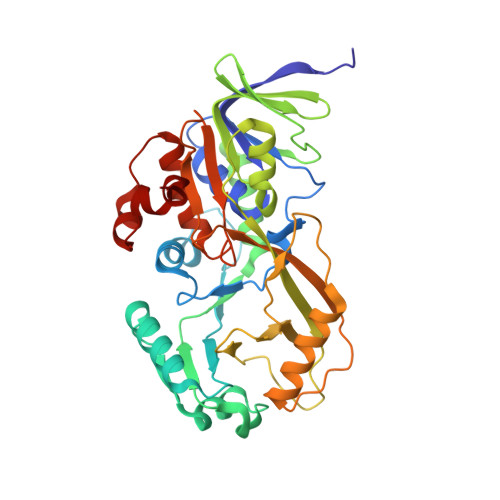
Conformational changes and substrate recognition in Pseudomonas aeruginosa D-arginine dehydrogenase.

Fu, G.Yuan, H.Li, C.Lu, C.D.Gadda, G.Weber, I.T.

(2010) Biochemistry 49: 8535-8545

  • DOI: https://doi.org/10.1021/bi1005865
  • Primary Citation of Related Structures:  
    3NYC, 3NYE, 3NYF

  • PubMed Abstract: 

    DADH catalyzes the flavin-dependent oxidative deamination of d-amino acids to the corresponding α-keto acids and ammonia. Here we report the first X-ray crystal structures of DADH at 1.06 Å resolution and its complexes with iminoarginine (DADH(red)/iminoarginine) and iminohistidine (DADH(red)/iminohistidine) at 1.30 Å resolution. The DADH crystal structure comprises an unliganded conformation and a product-bound conformation, which is almost identical to the DADH(red)/iminoarginine crystal structure. The active site of DADH was partially occupied with iminoarginine product (30% occupancy) that interacts with Tyr53 in the minor conformation of a surface loop. This flexible loop forms an "active site lid", similar to those seen in other enzymes, and may play an essential role in substrate recognition. The guanidinium side chain of iminoarginine forms a hydrogen bond interaction with the hydroxyl of Thr50 and an ionic interaction with Glu87. In the structure of DADH in complex with iminohistidine, two alternate conformations were observed for iminohistidine where the imidazole groups formed hydrogen bond interactions with the side chains of His48 and Thr50 and either Glu87 or Gln336. The different interactions and very distinct binding modes observed for iminoarginine and iminohistidine are consistent with the 1000-fold difference in k(cat)/K(m) values for d-arginine and d-histidine. Comparison of the kinetic data for the activity of DADH on different d-amino acids and the crystal structures in complex with iminoarginine and iminohistidine establishes that this enzyme is characterized by relatively broad substrate specificity, being able to oxidize positively charged and large hydrophobic d-amino acids bound within a flask-like cavity.


  • Organizational Affiliation

    Department of Biology, Georgia State University,Atlanta, Georgia 30303, USA.


Macromolecules
Find similar proteins by:  (by identity cutoff)  |  3D Structure
Entity ID: 1
MoleculeChains Sequence LengthOrganismDetailsImage
D-Arginine Dehydrogenase381Pseudomonas aeruginosaMutation(s): 0 
Gene Names: PA3863
EC: 1.4.1
UniProt
Find proteins for Q9HXE3 (Pseudomonas aeruginosa (strain ATCC 15692 / DSM 22644 / CIP 104116 / JCM 14847 / LMG 12228 / 1C / PRS 101 / PAO1))
Explore Q9HXE3 
Go to UniProtKB:  Q9HXE3
Entity Groups  
Sequence Clusters30% Identity50% Identity70% Identity90% Identity95% Identity100% Identity
UniProt GroupQ9HXE3
Sequence Annotations
Expand
  • Reference Sequence
Small Molecules
Ligands 2 Unique
IDChains Name / Formula / InChI Key2D Diagram3D Interactions
FAD
Query on FAD

Download Ideal Coordinates CCD File 
B [auth A]FLAVIN-ADENINE DINUCLEOTIDE
C27 H33 N9 O15 P2
VWWQXMAJTJZDQX-UYBVJOGSSA-N
IAR
Query on IAR

Download Ideal Coordinates CCD File 
C [auth A](2E)-5-[(diaminomethylidene)amino]-2-iminopentanoic acid
C6 H12 N4 O2
YWGYOCPWFDUKSA-QPJJXVBHSA-N
Experimental Data & Validation

Experimental Data

  • Method: X-RAY DIFFRACTION
  • Resolution: 1.30 Å
  • R-Value Free: 0.177 
  • R-Value Work: 0.143 
  • R-Value Observed: 0.143 
  • Space Group: P 21 21 21
Unit Cell:
Length ( Å )Angle ( ˚ )
a = 62.172α = 90
b = 78.428β = 90
c = 89.946γ = 90
Software Package:
Software NamePurpose
HKL-2000data collection
PHASERphasing
SHELXL-97refinement
HKL-2000data reduction
HKL-2000data scaling

Structure Validation

View Full Validation Report


Ligand Structure Quality Assessment 


Entry History 

Deposition Data

Revision History  (Full details and data files)

  • Version 1.0: 2010-09-15
    Type: Initial release
  • Version 1.1: 2011-07-13
    Changes: Version format compliance
  • Version 1.2: 2023-09-06
    Changes: Data collection, Database references, Derived calculations, Refinement description