3NFY

The Structure of Human Bisphosphoglycerate Mutase to 1.94A


Experimental Data Snapshot

  • Method: X-RAY DIFFRACTION
  • Resolution: 1.94 Å
  • R-Value Free: 0.277 
  • R-Value Work: 0.178 
  • R-Value Observed: 0.183 

wwPDB Validation   3D Report Full Report


This is version 1.2 of the entry. See complete history


Literature

Unliganded structure of human bisphosphoglycerate mutase reveals side-chain movements induced by ligand binding.

Patterson, A.Price, N.C.Nairn, J.

(2010) Acta Crystallogr Sect F Struct Biol Cryst Commun 66: 1415-1420

  • DOI: https://doi.org/10.1107/S1744309110035475
  • Primary Citation of Related Structures:  
    3NFY

  • PubMed Abstract: 

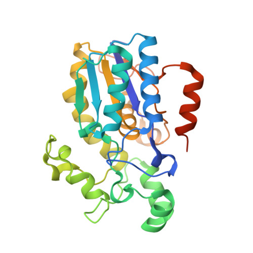
    Erythrocyte-specific bisphosphoglycerate mutase is a trifunctional enzyme which modulates the levels of 2,3-bisphosphoglycerate (2,3-BPG) in red blood cells by virtue of its synthase and phosphatase activities. Low levels of erythrocyte 2,3-BPG increase the affinity of haemoglobin for oxygen, thus limiting the release of oxygen into tissues. 2,3-BPG levels in stored blood decline rapidly owing to the phosphatase activity of bisphosphoglycerate mutase, which is enhanced by a fall in pH. Here, the 1.94 Å resolution X-ray structure of bisphosphoglycerate mutase is presented, focusing on the dynamic nature of key ligand-binding residues and their interaction with the inhibitor citrate. Residues at the binding pocket are complete. In addition, the movement of key residues in the presence and absence of ligand is described and alternative conformations are explored. The conformation in which the ligand citrate would bind at the substrate-binding pocket is proposed, with discussion and representations of its orientation. The characterization of bisphosphoglycerate mutase-citrate interactions will provide a framework for the design of specific inhibitors of the phosphatase activity of this enzyme, which may limit the decline of 2,3-BPG in stored blood.


  • Organizational Affiliation

    Division of Molecular and Cellular Biology, Institute of Biomedical and Life Sciences, University of Glasgow, Glasgow G12 8QQ, Scotland.


Macromolecules
Find similar proteins by:  (by identity cutoff)  |  3D Structure
Entity ID: 1
MoleculeChains Sequence LengthOrganismDetailsImage
Bisphosphoglycerate mutase
A, B
267Homo sapiensMutation(s): 0 
Gene Names: BPGM
EC: 5.4.2.4 (PDB Primary Data), 5.4.2.1 (PDB Primary Data), 3.1.3.13 (PDB Primary Data)
UniProt & NIH Common Fund Data Resources
Find proteins for P07738 (Homo sapiens)
Explore P07738 
Go to UniProtKB:  P07738
PHAROS:  P07738
GTEx:  ENSG00000172331 
Entity Groups  
Sequence Clusters30% Identity50% Identity70% Identity90% Identity95% Identity100% Identity
UniProt GroupP07738
Sequence Annotations
Expand
  • Reference Sequence
Experimental Data & Validation

Experimental Data

  • Method: X-RAY DIFFRACTION
  • Resolution: 1.94 Å
  • R-Value Free: 0.277 
  • R-Value Work: 0.178 
  • R-Value Observed: 0.183 
  • Space Group: P 1 21 1
Unit Cell:
Length ( Å )Angle ( ˚ )
a = 38.472α = 90
b = 61.345β = 95.87
c = 122.696γ = 90
Software Package:
Software NamePurpose
MOSFLMdata reduction
SCALAdata scaling
REFMACrefinement
PDB_EXTRACTdata extraction
ADSCdata collection

Structure Validation

View Full Validation Report


Entry History 

Deposition Data

Revision History  (Full details and data files)

  • Version 1.0: 2010-11-10
    Type: Initial release
  • Version 1.1: 2011-07-13
    Changes: Version format compliance
  • Version 1.2: 2023-09-06
    Changes: Data collection, Database references, Refinement description