


3N85
Crystallographic trimer of HER2 extracellular regions in complex with tryptophan-rich antibody fragment
- PDB DOI: https://doi.org/10.2210/pdb3N85/pdb
- Classification: Transferase/immune system
- Organism(s): Homo sapiens
- Expression System: Cricetulus griseus, Escherichia coli
- Mutation(s): No
- Deposited: 2010-05-27 Released: 2010-07-28
Experimental Data Snapshot
- Method: X-RAY DIFFRACTION
- Resolution: 3.20 Å
- R-Value Free: 0.230
- R-Value Work: 0.206
- R-Value Observed: 0.207
wwPDB Validation 3D Report Full Report

This is version 2.1 of the entry. See complete history.

Macromolecules
Find similar proteins by:
(by identity cutoff) | 3D Structure
Entity ID: 1 | |||||
|---|---|---|---|---|---|
| Molecule | Chains | Sequence Length | Organism | Details | Image |
| Receptor tyrosine-protein kinase erbB-2 | 624 | Homo sapiens | Mutation(s): 0 Gene Names: ERBB2, HER2, MLN19, NEU, NGL EC: 2.7.10.1 | 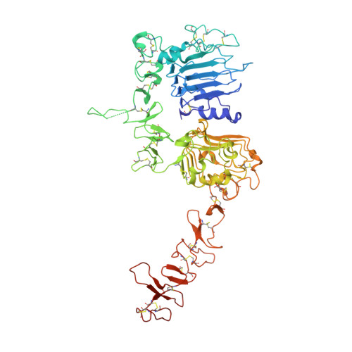 | |
UniProt & NIH Common Fund Data Resources | |||||
Find proteins for P04626 (Homo sapiens) Explore P04626 Go to UniProtKB: P04626 | |||||
PHAROS: P04626 GTEx: ENSG00000141736 | |||||
Entity Groups | |||||
| Sequence Clusters | 30% Identity50% Identity70% Identity90% Identity95% Identity100% Identity | ||||
| UniProt Group | P04626 | ||||
Sequence AnnotationsExpand | |||||
| |||||
Find similar proteins by:
(by identity cutoff) | 3D Structure
Entity ID: 2 | |||||
|---|---|---|---|---|---|
| Molecule | Chains | Sequence Length | Organism | Details | Image |
| Fab37 Light Chain | B [auth L] | 217 | Homo sapiens | Mutation(s): 0 | 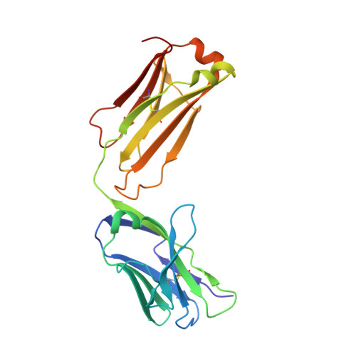 |
Entity Groups | |||||
| Sequence Clusters | 30% Identity50% Identity70% Identity90% Identity95% Identity100% Identity | ||||
Sequence AnnotationsExpand | |||||
| |||||
Find similar proteins by:
(by identity cutoff) | 3D Structure
Entity ID: 3 | |||||
|---|---|---|---|---|---|
| Molecule | Chains | Sequence Length | Organism | Details | Image |
| Fab37 Heavy Chain | C [auth H] | 224 | Homo sapiens | Mutation(s): 0 |  |
Entity Groups | |||||
| Sequence Clusters | 30% Identity50% Identity70% Identity90% Identity95% Identity100% Identity | ||||
Sequence AnnotationsExpand | |||||
| |||||
Oligosaccharides
Entity ID: 4 | |||||
|---|---|---|---|---|---|
| Molecule | Chains | Length | 2D Diagram | Glycosylation | 3D Interactions |
| beta-D-mannopyranose-(1-4)-2-acetamido-2-deoxy-beta-D-glucopyranose-(1-4)-[alpha-L-fucopyranose-(1-6)]2-acetamido-2-deoxy-beta-D-glucopyranose | D [auth B] | 4 |  | N-Glycosylation | |
Glycosylation Resources | |||||
GlyTouCan: G32152BH GlyCosmos: G32152BH GlyGen: G32152BH | |||||

Small Molecules
| Ligands 2 Unique | |||||
|---|---|---|---|---|---|
| ID | Chains | Name / Formula / InChI Key | 2D Diagram | 3D Interactions | |
| SO4 Query on SO4 | G [auth A] H [auth A] I [auth A] J [auth A] K [auth A] | SULFATE ION O4 S QAOWNCQODCNURD-UHFFFAOYSA-L |  | ||
| CL Query on CL | F [auth A] | CHLORIDE ION Cl VEXZGXHMUGYJMC-UHFFFAOYSA-M |  | ||
Experimental Data & Validation
Experimental Data
- Method: X-RAY DIFFRACTION
- Resolution: 3.20 Å
- R-Value Free: 0.230
- R-Value Work: 0.206
- R-Value Observed: 0.207
- Space Group: P 63 2 2
Unit Cell:
| Length ( Å ) | Angle ( ˚ ) |
|---|---|
| a = 182.22 | α = 90 |
| b = 182.22 | β = 90 |
| c = 330.851 | γ = 120 |
| Software Name | Purpose |
|---|---|
| Blu-Ice | data collection |
| PHASER | phasing |
| REFMAC | refinement |
| HKL-2000 | data reduction |
| SCALEPACK | data scaling |
Entry History
Deposition Data
- Released Date: 2010-07-28 Deposition Author(s): Eigenbrot, C.
Revision History (Full details and data files)
- Version 1.0: 2010-07-28
Type: Initial release - Version 1.1: 2011-07-13
Changes: Version format compliance - Version 1.2: 2014-01-15
Changes: Refinement description - Version 1.3: 2017-11-08
Changes: Refinement description - Version 1.4: 2018-01-24
Changes: Refinement description - Version 2.0: 2020-07-29
Type: Remediation
Reason: Carbohydrate remediation
Changes: Atomic model, Data collection, Derived calculations, Structure summary - Version 2.1: 2023-09-06
Changes: Data collection, Database references, Refinement description, Structure summary
