3N0I

Crystal Structure of Ad37 fiber knob in complex with GD1a oligosaccharide


Experimental Data Snapshot

  • Method: X-RAY DIFFRACTION
  • Resolution: 1.65 Å
  • R-Value Free: 0.187 
  • R-Value Work: 0.160 
  • R-Value Observed: 0.160 

wwPDB Validation   3D Report Full Report


This is version 2.1 of the entry. See complete history


Literature

The GD1a glycan is a cellular receptor for adenoviruses causing epidemic keratoconjunctivitis.

Nilsson, E.C.Storm, R.J.Bauer, J.Johansson, S.M.Lookene, A.Angstrom, J.Hedenstrom, M.Eriksson, T.L.Frangsmyr, L.Rinaldi, S.Willison, H.J.Domellof, F.P.Stehle, T.Arnberg, N.

(2011) Nat Med 17: 105-109

  • DOI: https://doi.org/10.1038/nm.2267
  • Primary Citation of Related Structures:  
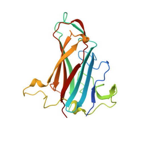
    3N0I

  • PubMed Abstract: 

    Adenovirus type 37 (Ad37) is a leading cause of epidemic keratoconjunctivitis (EKC), a severe and highly contagious ocular disease. Whereas most other adenoviruses infect cells by engaging CD46 or the coxsackie and adenovirus receptor (CAR), Ad37 binds previously unknown sialic acid-containing cell surface molecules. By glycan array screening, we show here that the receptor-recognizing knob domain of the Ad37 fiber protein specifically binds a branched hexasaccharide that is present in the GD1a ganglioside and that features two terminal sialic acids. Soluble GD1a glycan and GD1a-binding antibodies efficiently prevented Ad37 virions from binding and infecting corneal cells. Unexpectedly, the receptor is constituted by one or more glycoproteins containing the GD1a glycan motif rather than the ganglioside itself, as shown by binding, infection and flow cytometry experiments. Molecular modeling, nuclear magnetic resonance and X-ray crystallography reveal that the two terminal sialic acids dock into two of three previously established sialic acid-binding sites in the trimeric Ad37 knob. Surface plasmon resonance analysis shows that the knob-GD1a glycan interaction has high affinity. Our findings therefore form a basis for the design and development of sialic acid-containing antiviral drugs for topical treatment of EKC.


  • Organizational Affiliation

    Department of Clinical Microbiology, Division of Virology, Umeå University, Umeå, Sweden.


Macromolecules
Find similar proteins by:  (by identity cutoff)  |  3D Structure
Entity ID: 1
MoleculeChains Sequence LengthOrganismDetailsImage
Fiber
A, B, C
194Human adenovirus D37Mutation(s): 0 
Gene Names: L5
UniProt
Find proteins for Q64823 (Human adenovirus D37)
Explore Q64823 
Go to UniProtKB:  Q64823
Entity Groups  
Sequence Clusters30% Identity50% Identity70% Identity90% Identity95% Identity100% Identity
UniProt GroupQ64823
Sequence Annotations
Expand
  • Reference Sequence
Oligosaccharides

Help

Entity ID: 2
MoleculeChains Length2D Diagram Glycosylation3D Interactions
N-acetyl-alpha-neuraminic acid-(2-3)-beta-D-galactopyranose-(1-3)-2-acetamido-2-deoxy-beta-D-galactopyranose-(1-4)-[N-acetyl-alpha-neuraminic acid-(2-3)]beta-D-galactopyranose-(1-4)-beta-D-glucopyranose
D
6N/A
Glycosylation Resources
GlyTouCan:  G46677TE
GlyCosmos:  G46677TE
GlyGen:  G46677TE
Experimental Data & Validation

Experimental Data

  • Method: X-RAY DIFFRACTION
  • Resolution: 1.65 Å
  • R-Value Free: 0.187 
  • R-Value Work: 0.160 
  • R-Value Observed: 0.160 
  • Space Group: P 1 21 1
Unit Cell:
Length ( Å )Angle ( ˚ )
a = 60.99α = 90
b = 69.77β = 94.92
c = 74.9γ = 90
Software Package:
Software NamePurpose
XDSdata scaling
PHASERphasing
REFMACrefinement
XDSdata reduction

Structure Validation

View Full Validation Report


Entry History 

Revision History  (Full details and data files)

  • Version 1.0: 2010-12-29
    Type: Initial release
  • Version 1.1: 2011-07-13
    Changes: Version format compliance
  • Version 1.2: 2014-02-19
    Changes: Database references
  • Version 2.0: 2020-07-29
    Type: Remediation
    Reason: Carbohydrate remediation
    Changes: Atomic model, Data collection, Database references, Derived calculations, Structure summary
  • Version 2.1: 2023-09-06
    Changes: Data collection, Database references, Refinement description, Structure summary