3MQH

crystal structure of the 3-N-acetyl transferase WlbB from Bordetella petrii in complex with CoA and UDP-3-amino-2-acetamido-2,3-dideoxy glucuronic acid


Experimental Data Snapshot

  • Method: X-RAY DIFFRACTION
  • Resolution: 1.43 Å
  • R-Value Free: 0.206 
  • R-Value Work: 0.174 
  • R-Value Observed: 0.176 

wwPDB Validation   3D Report Full Report


Ligand Structure Quality Assessment 


This is version 1.2 of the entry. See complete history


Literature

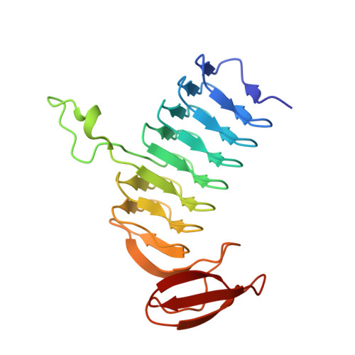
Molecular structure of WlbB, a bacterial N-acetyltransferase involved in the biosynthesis of 2,3-diacetamido-2,3-dideoxy-D-mannuronic acid .

Thoden, J.B.Holden, H.M.

(2010) Biochemistry 49: 4644-4653

  • DOI: https://doi.org/10.1021/bi1005738
  • Primary Citation of Related Structures:  
    3MQG, 3MQH

  • PubMed Abstract: 

    The pathogenic bacteria Pseudomonas aeruginosa and Bordetella pertussis contain in their outer membranes the rare sugar 2,3-diacetamido-2,3-dideoxy-d-mannuronic acid. Five enzymes are required for the biosynthesis of this sugar starting from UDP-N-acetylglucosamine. One of these, referred to as WlbB, is an N-acetyltransferase that converts UDP-2-acetamido-3-amino-2,3-dideoxy-d-glucuronic acid (UDP-GlcNAc3NA) to UDP-2,3-diacetamido-2,3-dideoxy-d-glucuronic acid (UDP-GlcNAc3NAcA). Here we report the three-dimensional structure of WlbB from Bordetella petrii. For this analysis, two ternary structures were determined to 1.43 A resolution: one in which the protein was complexed with acetyl-CoA and UDP and the second in which the protein contained bound CoA and UDP-GlcNAc3NA. WlbB adopts a trimeric quaternary structure and belongs to the LbetaH superfamily of N-acyltransferases. Each subunit contains 27 beta-strands, 23 of which form the canonical left-handed beta-helix. There are only two hydrogen bonds that occur between the protein and the GlcNAc3NA moiety, one between O(delta1) of Asn 84 and the sugar C-3' amino group and the second between the backbone amide group of Arg 94 and the sugar C-5' carboxylate. The sugar C-3' amino group is ideally positioned in the active site to attack the si face of acetyl-CoA. Given that there are no protein side chains that can function as general bases within the GlcNAc3NA binding pocket, a reaction mechanism is proposed for WlbB whereby the sulfur of CoA ultimately functions as the proton acceptor required for catalysis.


  • Organizational Affiliation

    Department of Biochemistry, University of Wisconsin, Madison, Wisconsin 53706, USA.


Macromolecules
Find similar proteins by:  (by identity cutoff)  |  3D Structure
Entity ID: 1
MoleculeChains Sequence LengthOrganismDetailsImage
Lipopolysaccharides biosynthesis acetyltransferase
A, B, C, D, E
A, B, C, D, E, F
192Bordetella petrii DSM 12804Mutation(s): 0 
Gene Names: Bpet4817wlbB
EC: 2.3.1
UniProt
Find proteins for A9IH93 (Bordetella petrii (strain ATCC BAA-461 / DSM 12804 / CCUG 43448))
Explore A9IH93 
Go to UniProtKB:  A9IH93
Entity Groups  
Sequence Clusters30% Identity50% Identity70% Identity90% Identity95% Identity100% Identity
UniProt GroupA9IH93
Sequence Annotations
Expand
  • Reference Sequence
Small Molecules
Ligands 6 Unique
IDChains Name / Formula / InChI Key2D Diagram3D Interactions
COA
Query on COA

Download Ideal Coordinates CCD File 
DA [auth E]
FA [auth F]
H [auth A]
M [auth B]
P [auth C]
DA [auth E],
FA [auth F],
H [auth A],
M [auth B],
P [auth C],
X [auth D]
COENZYME A
C21 H36 N7 O16 P3 S
RGJOEKWQDUBAIZ-IBOSZNHHSA-N
MJZ
Query on MJZ

Download Ideal Coordinates CCD File 
CA [auth E]
G [auth A]
GA [auth F]
L [auth B]
Q [auth C]
CA [auth E],
G [auth A],
GA [auth F],
L [auth B],
Q [auth C],
W [auth D]
(2S,3S,4R,5R,6R)-5-(acetylamino)-4-amino-6-{[(R)-{[(R)-{[(2R,3S,4R,5R)-5-(2,4-dioxo-3,4-dihydropyrimidin-1(2H)-yl)-3,4-dihydroxytetrahydrofuran-2-yl]methoxy}(hydroxy)phosphoryl]oxy}(hydroxy)phosphoryl]oxy}-3-hydroxytetrahydro-2H-pyran-2-carboxylic acid
C17 H26 N4 O17 P2
RRAQYLXLCYIZBB-HHKCBAECSA-N
PE4
Query on PE4

Download Ideal Coordinates CCD File 
V [auth C]2-{2-[2-(2-{2-[2-(2-ETHOXY-ETHOXY)-ETHOXY]-ETHOXY}-ETHOXY)-ETHOXY]-ETHOXY}-ETHANOL
C16 H34 O8
PJWQOENWHPEPKI-UHFFFAOYSA-N
PO4
Query on PO4

Download Ideal Coordinates CCD File 
HA [auth F],
R [auth C]
PHOSPHATE ION
O4 P
NBIIXXVUZAFLBC-UHFFFAOYSA-K
EDO
Query on EDO

Download Ideal Coordinates CCD File 
AA [auth D]
EA [auth E]
I [auth A]
IA [auth F]
K [auth A]
AA [auth D],
EA [auth E],
I [auth A],
IA [auth F],
K [auth A],
N [auth B],
O [auth B],
S [auth C],
T [auth C],
U [auth C],
Y [auth D],
Z [auth D]
1,2-ETHANEDIOL
C2 H6 O2
LYCAIKOWRPUZTN-UHFFFAOYSA-N
NA
Query on NA

Download Ideal Coordinates CCD File 
BA [auth D],
J [auth A]
SODIUM ION
Na
FKNQFGJONOIPTF-UHFFFAOYSA-N
Experimental Data & Validation

Experimental Data

  • Method: X-RAY DIFFRACTION
  • Resolution: 1.43 Å
  • R-Value Free: 0.206 
  • R-Value Work: 0.174 
  • R-Value Observed: 0.176 
  • Space Group: P 1 21 1
Unit Cell:
Length ( Å )Angle ( ˚ )
a = 68.466α = 90
b = 107.891β = 102.55
c = 91.269γ = 90
Software Package:
Software NamePurpose
ADSCdata collection
PHASERphasing
REFMACrefinement
HKL-3000data reduction
HKL-3000data scaling

Structure Validation

View Full Validation Report


Ligand Structure Quality Assessment 


Entry History 

Deposition Data

Revision History  (Full details and data files)

  • Version 1.0: 2010-05-12
    Type: Initial release
  • Version 1.1: 2011-07-13
    Changes: Version format compliance
  • Version 1.2: 2023-09-06
    Changes: Data collection, Database references, Derived calculations, Refinement description