Structural origins of pH-dependent chemical shifts in the B1 domain of protein G.
Tomlinson, J.H., Green, V.L., Baker, P.J., Williamson, M.P.(2010) Proteins 78: 3000-3016
- PubMed: 20715051
- DOI: https://doi.org/10.1002/prot.22825
- Primary Citation of Related Structures:
3MP9 - PubMed Abstract:
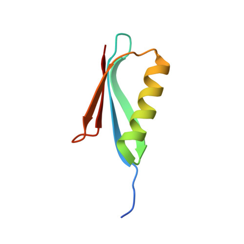
We report chemical shifts for H(N), N, and C' nuclei in the His-tagged B1 domain of protein G (GB1) over a range of pH values from pH 2.0 to 9.0, which fit well to standard pH-dependent equations. We also report a 1.2 Å resolution crystal structure of GB1 at pH 3.0. Comparison of this crystal structure with published crystal structures at higher pHs provides details of the structural changes in GB1 associated with protonation of the carboxylate groups, in particular a conformational change in the C-terminus of the protein at low pH. An additional change described recently is not seen in the crystal structure because of crystal contacts. We show that the pH-dependent changes in chemical shifts can be almost entirely understood based on structural changes, thereby providing insight into the relationship between structure and chemical shift. In particular, we describe through-bond effects extending up to five bonds, affecting N and C' but not H(N); through-space effects of carboxylates, which fit well to a simple electric field model; and effects due to conformational change, which have a similar magnitude to many of the direct effects. Finally, we discuss cooperative effects, demonstrating a lack of cooperative unfolding in the helix, and the existence of a β-sheet "iceberg" extending over three of the four strands. This study therefore extends the application of chemical shifts to understanding protein structure.
Organizational Affiliation:
Department of Molecular Biology and Biotechnology, University of Sheffield, Firth Court, Western Bank, Sheffield S10 2TN, United Kingdom.