3MP3

Crystal Structure of Human Lyase in complex with inhibitor HG-CoA


Experimental Data Snapshot

  • Method: X-RAY DIFFRACTION
  • Resolution: 2.40 Å
  • R-Value Free: 0.281 
  • R-Value Work: 0.212 
  • R-Value Observed: 0.212 

wwPDB Validation   3D Report Full Report


Ligand Structure Quality Assessment 


This is version 1.2 of the entry. See complete history


Literature

Functional insights into human HMG-CoA lyase from structures of Acyl-CoA-containing ternary complexes.

Fu, Z.Runquist, J.A.Montgomery, C.Miziorko, H.M.Kim, J.J.

(2010) J Biol Chem 285: 26341-26349

  • DOI: https://doi.org/10.1074/jbc.M110.139931
  • Primary Citation of Related Structures:  
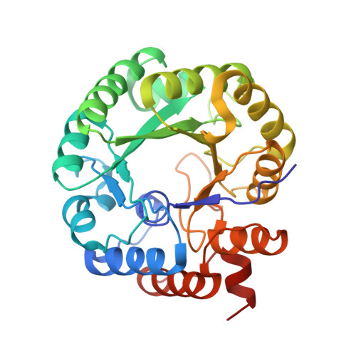
    3MP3, 3MP4, 3MP5

  • PubMed Abstract: 

    HMG-CoA lyase (HMGCL) is crucial to ketogenesis, and inherited human mutations are potentially lethal. Detailed understanding of the HMGCL reaction mechanism and the molecular basis for correlating human mutations with enzyme deficiency have been limited by the lack of structural information for enzyme liganded to an acyl-CoA substrate or inhibitor. Crystal structures of ternary complexes of WT HMGCL with the competitive inhibitor 3-hydroxyglutaryl-CoA and of the catalytically deficient HMGCL R41M mutant with substrate HMG-CoA have been determined to 2.4 and 2.2 A, respectively. Comparison of these beta/alpha-barrel structures with those of unliganded HMGCL and R41M reveals substantial differences for Mg(2+) coordination and positioning of the flexible loop containing the conserved HMGCL "signature" sequence. In the R41M-Mg(2+)-substrate ternary complex, loop residue Cys(266) (implicated in active-site function by mechanistic and mutagenesis observations) is more closely juxtaposed to the catalytic site than in the case of unliganded enzyme or the WT enzyme-Mg(2+)-3-hydroxyglutaryl-CoA inhibitor complex. In both ternary complexes, the S-stereoisomer of substrate or inhibitor is specifically bound, in accord with the observed Mg(2+) liganding of both C3 hydroxyl and C5 carboxyl oxygens. In addition to His(233) and His(235) imidazoles, other Mg(2+) ligands are the Asp(42) carboxyl oxygen and an ordered water molecule. This water, positioned between Asp(42) and the C3 hydroxyl of bound substrate/inhibitor, may function as a proton shuttle. The observed interaction of Arg(41) with the acyl-CoA C1 carbonyl oxygen explains the effects of Arg(41) mutation on reaction product enolization and explains why human Arg(41) mutations cause drastic enzyme deficiency.


  • Organizational Affiliation

    Department of Biochemistry, Medical College of Wisconsin, Milwaukee, Wisconsin 53226, USA.


Macromolecules
Find similar proteins by:  (by identity cutoff)  |  3D Structure
Entity ID: 1
MoleculeChains Sequence LengthOrganismDetailsImage
Hydroxymethylglutaryl-CoA lyase
A, B, C, D, E
A, B, C, D, E, F
298Homo sapiensMutation(s): 0 
Gene Names: HMGCL
EC: 4.1.3.4
UniProt & NIH Common Fund Data Resources
Find proteins for P35914 (Homo sapiens)
Explore P35914 
Go to UniProtKB:  P35914
PHAROS:  P35914
GTEx:  ENSG00000117305 
Entity Groups  
Sequence Clusters30% Identity50% Identity70% Identity90% Identity95% Identity100% Identity
UniProt GroupP35914
Sequence Annotations
Expand
  • Reference Sequence
Small Molecules
Ligands 3 Unique
IDChains Name / Formula / InChI Key2D Diagram3D Interactions
HGG
Query on HGG

Download Ideal Coordinates CCD File 
I [auth B](3R,5S,9R,21S)-1-[(2R,3S,4R,5R)-5-(6-amino-9H-purin-9-yl)-4-hydroxy-3-(phosphonooxy)tetrahydrofuran-2-yl]-3,5,9,21-tetrahydroxy-8,8-dimethyl-10,14,19-trioxo-2,4,6-trioxa-18-thia-11,15-diaza-3,5-diphosphatricosan-23-oic acid 3,5-dioxide
C26 H42 N7 O20 P3 S
IIYZSYKTQRIPRG-UPWLPGGHSA-N
3HG
Query on 3HG

Download Ideal Coordinates CCD File 
G [auth A]3-HYDROXYPENTANEDIOIC ACID
C5 H8 O5
ZQHYXNSQOIDNTL-UHFFFAOYSA-N
MG
Query on MG

Download Ideal Coordinates CCD File 
H [auth A]
J [auth B]
K [auth C]
L [auth D]
M [auth E]
H [auth A],
J [auth B],
K [auth C],
L [auth D],
M [auth E],
N [auth F]
MAGNESIUM ION
Mg
JLVVSXFLKOJNIY-UHFFFAOYSA-N
Experimental Data & Validation

Experimental Data

  • Method: X-RAY DIFFRACTION
  • Resolution: 2.40 Å
  • R-Value Free: 0.281 
  • R-Value Work: 0.212 
  • R-Value Observed: 0.212 
  • Space Group: C 1 2 1
Unit Cell:
Length ( Å )Angle ( ˚ )
a = 197.37α = 90
b = 117β = 112.31
c = 86.87γ = 90
Software Package:
Software NamePurpose
CrystalCleardata collection
CCP4model building
CNSrefinement
HKL-2000data reduction
SCALAdata scaling
CCP4phasing

Structure Validation

View Full Validation Report


Ligand Structure Quality Assessment 


Entry History 

Deposition Data

Revision History  (Full details and data files)

  • Version 1.0: 2010-06-16
    Type: Initial release
  • Version 1.1: 2011-07-13
    Changes: Version format compliance
  • Version 1.2: 2023-09-06
    Changes: Data collection, Database references, Derived calculations, Refinement description