3MKJ

Methionine gamma-lyase from Citrobacter freundii with pyridoximine-5'-phosphate


Experimental Data Snapshot

  • Method: X-RAY DIFFRACTION
  • Resolution: 1.65 Å
  • R-Value Free: 0.232 
  • R-Value Work: 0.196 
  • R-Value Observed: 0.198 

wwPDB Validation   3D Report Full Report


Ligand Structure Quality Assessment 


This is version 1.4 of the entry. See complete history


Literature

Exploring methionine gamma-lyase structure-function relationship via microspectrophotometry and X-ray crystallography

Ronda, L.Bazhulina, N.P.Morozova, E.A.Revtovich, S.V.Chekhov, V.O.Nikulin, A.D.Demidkina, T.V.Mozzarelli, A.

(2011) Biochim Biophys Acta 1814: 834-842

  • DOI: https://doi.org/10.1016/j.bbapap.2010.06.017
  • Primary Citation of Related Structures:  
    3MKJ

  • PubMed Abstract: 

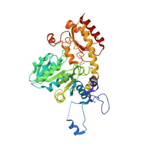
    Pyridoxal 5'-phosphate (PLP) dependent methionine γ-lyase catalyzes the breakdown of L-methionine to α-ketobutyric acid, methanethiol and ammonia. This enzyme, present in anaerobic microorganisms, has biomedical interest both for its activity as antitumor agent, depleting methionine supply in methionine-dependent cancers, and as target in the treatment of human pathogen infections, activating the pro-drug trifluoromethionine. To validate the structure of the enzyme from Citrobacter freundii, crystallized from monomethyl ether polyethylene glycol 2000, for the development of lead compounds, the reactivity of the crystalline enzyme towards L-methionine, substrate analogs and inhibitors was determined by polarized absorption microspectrophotometry. Spectral data were also collected for enzyme crystals, grown in monomethyl ether polyethylene glycol 2000 in the presence of ammonium sulfate. The three-dimensional structure of these enzyme crystals, solved at 1.65Å resolution with R(free) 23.2%, revealed the surprising absence of the aldimine bond between the active site Lys210 and PLP. Different hypothesis are proposed and discussed in the light of spectral and structural data, pointing out to the relevance of the complementarity between X-ray crystallography and single crystal spectroscopy for the understanding of biological mechanisms at molecular level. This article is part of a Special Issue entitled: Protein Structure and Function in the Crystalline State.


  • Organizational Affiliation

    Department of Biochemistry and Molecular Biology, University of Parma, Parma, Italy.


Macromolecules
Find similar proteins by:  (by identity cutoff)  |  3D Structure
Entity ID: 1
MoleculeChains Sequence LengthOrganismDetailsImage
Methionine gamma-lyase398Citrobacter freundiiMutation(s): 0 
Gene Names: megL
EC: 4.4.1.11
UniProt
Find proteins for Q84AR1 (Citrobacter freundii)
Explore Q84AR1 
Go to UniProtKB:  Q84AR1
Entity Groups  
Sequence Clusters30% Identity50% Identity70% Identity90% Identity95% Identity100% Identity
UniProt GroupQ84AR1
Sequence Annotations
Expand
  • Reference Sequence
Small Molecules
Ligands 1 Unique
IDChains Name / Formula / InChI Key2D Diagram3D Interactions
PZP
Query on PZP

Download Ideal Coordinates CCD File 
B [auth A][5-hydroxy-4-(iminomethyl)-6-methyl-pyridin-3-yl]methyl dihydrogen phosphate
C8 H11 N2 O5 P
CLOXDHRIVOURLQ-XNWCZRBMSA-N
Modified Residues  1 Unique
IDChains TypeFormula2D DiagramParent
CSO
Query on CSO
A
L-PEPTIDE LINKINGC3 H7 N O3 SCYS
Experimental Data & Validation

Experimental Data

  • Method: X-RAY DIFFRACTION
  • Resolution: 1.65 Å
  • R-Value Free: 0.232 
  • R-Value Work: 0.196 
  • R-Value Observed: 0.198 
  • Space Group: I 2 2 2
Unit Cell:
Length ( Å )Angle ( ˚ )
a = 56.69α = 90
b = 122.8β = 90
c = 128.04γ = 90
Software Package:
Software NamePurpose
MAR345dtbdata collection
MOLREPphasing
PHENIXrefinement
XDSdata reduction
XDSdata scaling

Structure Validation

View Full Validation Report


Ligand Structure Quality Assessment 


Entry History 

Deposition Data

Revision History  (Full details and data files)

  • Version 1.0: 2010-08-11
    Type: Initial release
  • Version 1.1: 2011-07-13
    Changes: Version format compliance
  • Version 1.2: 2014-03-05
    Changes: Database references
  • Version 1.3: 2023-11-01
    Changes: Data collection, Database references, Derived calculations, Refinement description
  • Version 1.4: 2023-11-22
    Changes: Data collection