3KWY

Crystal structure of RXRalpha ligand binding domain in complex with triphenyltin and a coactivator fragment


Experimental Data Snapshot

  • Method: X-RAY DIFFRACTION
  • Resolution: 2.30 Å
  • R-Value Free: 0.244 
  • R-Value Work: 0.183 
  • R-Value Observed: 0.186 

wwPDB Validation   3D Report Full Report


Ligand Structure Quality Assessment 


This is version 1.3 of the entry. See complete history


Literature

A structural view of nuclear hormone receptor: endocrine disruptor interactions.

le Maire, A.Bourguet, W.Balaguer, P.

(2010) Cell Mol Life Sci 67: 1219-1237

  • DOI: https://doi.org/10.1007/s00018-009-0249-2
  • Primary Citation of Related Structures:  
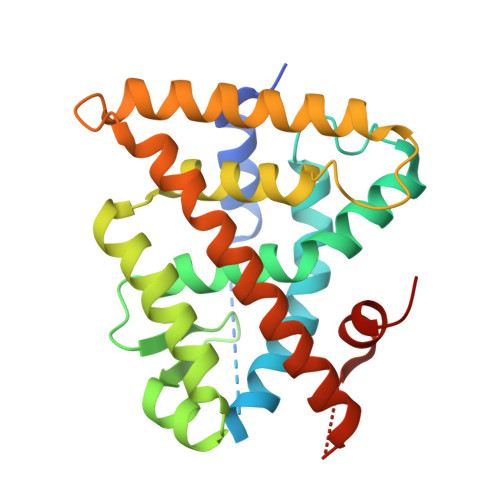
    3KWY

  • PubMed Abstract: 

    Endocrine-disrupting chemicals (EDCs) represent a broad class of exogenous substances that cause adverse effects in the endocrine system by interfering with hormone biosynthesis, metabolism, or action. The molecular mechanisms of EDCs involve different pathways including interactions with nuclear hormone receptors (NHRs) which are primary targets of a large variety of environmental contaminants. Here, based on the crystal structures currently available in the Protein Data Bank, we review recent studies showing the many ways in which EDCs interact with NHRs and impact their signaling pathways. Like the estrogenic chemical diethylstilbestrol, some EDCs mimic the natural hormones through conserved protein-ligand contacts, while others, such as organotins, employ radically different binding mechanisms. Such structure-based knowledge, in addition to providing a better understanding of EDC activities, can be used to predict the endocrine-disrupting potential of environmental pollutants and may have applications in drug discovery.


  • Organizational Affiliation

    INSERM, U554, Centre de Biochimie Structurale, Montpellier, France.


Macromolecules
Find similar proteins by:  (by identity cutoff)  |  3D Structure
Entity ID: 1
MoleculeChains Sequence LengthOrganismDetailsImage
Retinoic acid receptor RXR-alpha244Homo sapiensMutation(s): 0 
Gene Names: RXRANR2B1
UniProt & NIH Common Fund Data Resources
Find proteins for P19793 (Homo sapiens)
Explore P19793 
Go to UniProtKB:  P19793
PHAROS:  P19793
GTEx:  ENSG00000186350 
Entity Groups  
Sequence Clusters30% Identity50% Identity70% Identity90% Identity95% Identity100% Identity
UniProt GroupP19793
Sequence Annotations
Expand
  • Reference Sequence

Find similar proteins by:  Sequence   |   3D Structure  

Entity ID: 2
MoleculeChains Sequence LengthOrganismDetailsImage
Nuclear receptor coactivator 2 peptide13Homo sapiensMutation(s): 0 
Gene Names: NCOA2
UniProt & NIH Common Fund Data Resources
Find proteins for Q15596 (Homo sapiens)
Explore Q15596 
Go to UniProtKB:  Q15596
PHAROS:  Q15596
GTEx:  ENSG00000140396 
Entity Groups  
Sequence Clusters30% Identity50% Identity70% Identity90% Identity95% Identity100% Identity
UniProt GroupQ15596
Sequence Annotations
Expand
  • Reference Sequence
Small Molecules
Experimental Data & Validation

Experimental Data

  • Method: X-RAY DIFFRACTION
  • Resolution: 2.30 Å
  • R-Value Free: 0.244 
  • R-Value Work: 0.183 
  • R-Value Observed: 0.186 
  • Space Group: P 43 21 2
Unit Cell:
Length ( Å )Angle ( ˚ )
a = 64.635α = 90
b = 64.635β = 90
c = 113.589γ = 90
Software Package:
Software NamePurpose
ADSCdata collection
REFMACrefinement
MOSFLMdata reduction
SCALAdata scaling
REFMACphasing

Structure Validation

View Full Validation Report


Ligand Structure Quality Assessment 


Entry History 

Deposition Data

Revision History  (Full details and data files)

  • Version 1.0: 2010-01-19
    Type: Initial release
  • Version 1.1: 2011-07-13
    Changes: Advisory, Refinement description, Version format compliance
  • Version 1.2: 2022-08-17
    Changes: Database references, Derived calculations
  • Version 1.3: 2023-09-06
    Changes: Data collection, Refinement description