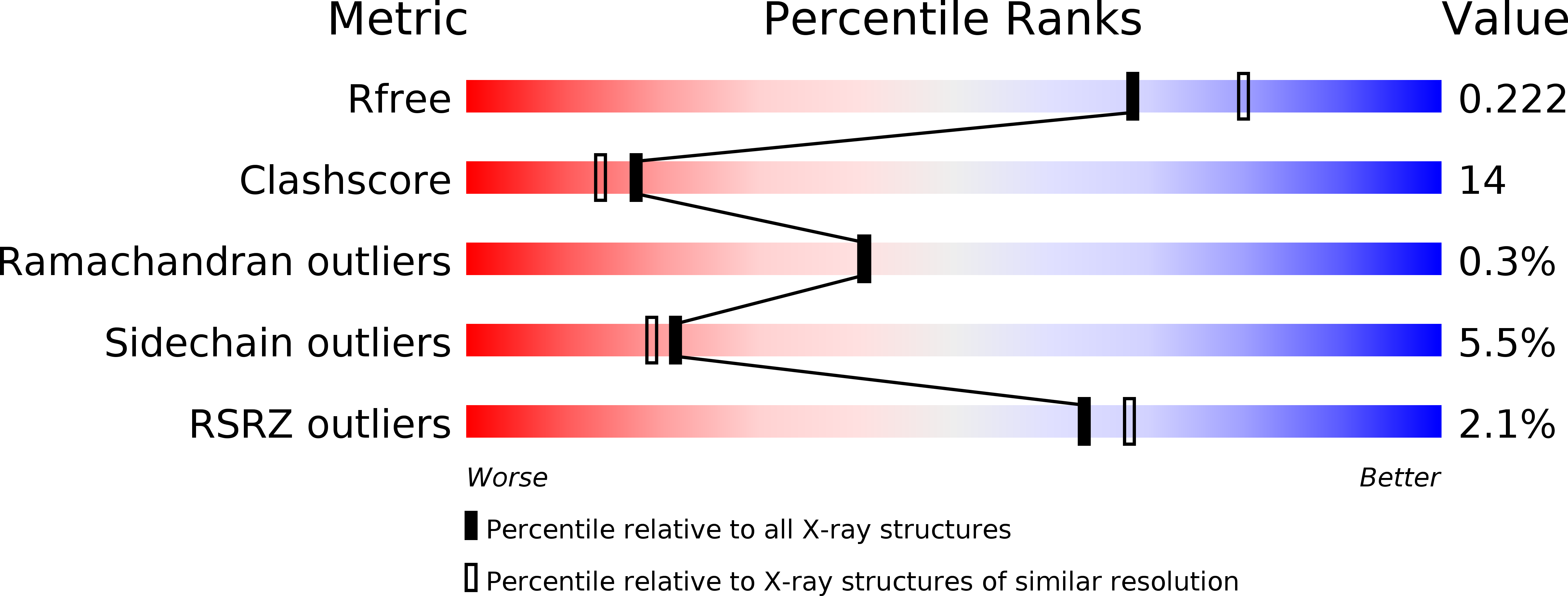
Glycal formation in crystals of uridine phosphorylase.
Paul, D., O'Leary, S.E., Rajashankar, K., Bu, W., Toms, A., Settembre, E.C., Sanders, J.M., Begley, T.P., Ealick, S.E.(2010) Biochemistry 49: 3499-3509
- PubMed: 20364833
- DOI: https://doi.org/10.1021/bi902073b
- Primary Citation of Related Structures:
3KU4, 3KUK, 3KVR, 3KVV, 3KVY - PubMed Abstract:
Uridine phosphorylase is a key enzyme in the pyrimidine salvage pathway. This enzyme catalyzes the reversible phosphorolysis of uridine to uracil and ribose 1-phosphate (or 2'-deoxyuridine to 2'-deoxyribose 1-phosphate). Here we report the structure of hexameric Escherichia coli uridine phosphorylase treated with 5-fluorouridine and sulfate and dimeric bovine uridine phosphorylase treated with 5-fluoro-2'-deoxyuridine or uridine, plus sulfate. In each case the electron density shows three separate species corresponding to the pyrimidine base, sulfate, and a ribosyl species, which can be modeled as a glycal. In the structures of the glycal complexes, the fluorouracil O2 atom is appropriately positioned to act as the base required for glycal formation via deprotonation at C2'. Crystals of bovine uridine phosphorylase treated with 2'-deoxyuridine and sulfate show intact nucleoside. NMR time course studies demonstrate that uridine phosphorylase can catalyze the hydrolysis of the fluorinated nucleosides in the absence of phosphate or sulfate, without the release of intermediates or enzyme inactivation. These results add a previously unencountered mechanistic motif to the body of information on glycal formation by enzymes catalyzing the cleavage of glycosyl bonds.
Organizational Affiliation:
Department of Chemistry and Chemical Biology, Cornell University, Ithaca, New York 14853-1301, USA.


