


3KU0
Structure of GAP31 with adenine at its binding pocket
- PDB DOI: https://doi.org/10.2210/pdb3KU0/pdb
- Classification: HYDROLASE
- Organism(s): Suregada multiflora
- Mutation(s): No
- Deposited: 2009-11-26 Released: 2010-01-26
Experimental Data Snapshot
- Method: X-RAY DIFFRACTION
- Resolution: 1.90 Å
- R-Value Free: 0.242
- R-Value Work: 0.200
- R-Value Observed: 0.200

This is version 1.3 of the entry. See complete history.

Macromolecules
Find similar proteins by:
(by identity cutoff) | 3D Structure
Entity ID: 1 | |||||
|---|---|---|---|---|---|
| Molecule | Chains | Sequence Length | Organism | Details | Image |
| Ribosome-inactivating protein gelonin | 251 | Suregada multiflora | Mutation(s): 0 EC: 3.2.2.22 | 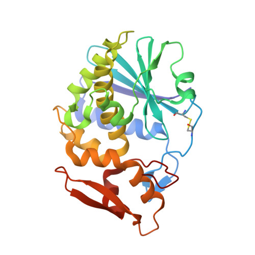 | |
UniProt | |||||
Find proteins for P33186 (Suregada multiflora) Explore P33186 Go to UniProtKB: P33186 | |||||
Entity Groups | |||||
| Sequence Clusters | 30% Identity50% Identity70% Identity90% Identity95% Identity100% Identity | ||||
| UniProt Group | P33186 | ||||
Sequence AnnotationsExpand | |||||
| |||||
Small Molecules
| Ligands 2 Unique | |||||
|---|---|---|---|---|---|
| ID | Chains | Name / Formula / InChI Key | 2D Diagram | 3D Interactions | |
| NAG Query on NAG | D [auth A], F [auth B] | 2-acetamido-2-deoxy-beta-D-glucopyranose C8 H15 N O6 OVRNDRQMDRJTHS-FMDGEEDCSA-N |  | ||
| ADE Query on ADE | C [auth A], E [auth B] | ADENINE C5 H5 N5 GFFGJBXGBJISGV-UHFFFAOYSA-N |  | ||
Experimental Data & Validation
Experimental Data
- Method: X-RAY DIFFRACTION
- Resolution: 1.90 Å
- R-Value Free: 0.242
- R-Value Work: 0.200
- R-Value Observed: 0.200
- Space Group: P 1 21 1
Unit Cell:
| Length ( Å ) | Angle ( ˚ ) |
|---|---|
| a = 48.373 | α = 90 |
| b = 44.395 | β = 98.38 |
| c = 137.27 | γ = 90 |
| Software Name | Purpose |
|---|---|
| CNS | refinement |
| PDB_EXTRACT | data extraction |
| HKL-2000 | data collection |
| HKL-2000 | data reduction |
| HKL-2000 | data scaling |
| CNS | phasing |
Entry History
Deposition Data
- Released Date: 2010-01-26 Deposition Author(s): Kong, X.-P.
Revision History (Full details and data files)
- Version 1.0: 2010-01-26
Type: Initial release - Version 1.1: 2011-07-13
Changes: Version format compliance - Version 1.2: 2020-02-26
Changes: Data collection, Derived calculations - Version 1.3: 2020-07-29
Type: Remediation
Reason: Carbohydrate remediation
Changes: Data collection, Derived calculations, Structure summary
