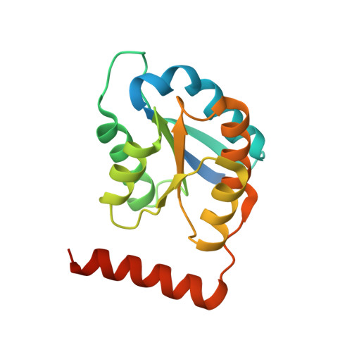
Structures of type B ribose 5-phosphate isomerase from Trypanosoma cruzi shed light on the determinants of sugar specificity in the structural family.
Stern, A.L., Naworyta, A., Cazzulo, J.J., Mowbray, S.L.(2011) FEBS J 278: 793-808
- PubMed: 21205211
- DOI: https://doi.org/10.1111/j.1742-4658.2010.07999.x
- Primary Citation of Related Structures:
3K7O, 3K7P, 3K7S, 3K8C, 3M1P - PubMed Abstract:
Ribose-5-phosphate isomerase (Rpi; EC 5.3.1.6) is a key activity of the pentose phosphate pathway. Two unrelated types of sequence/structure possess this activity: type A Rpi (present in most organisms) and type B Rpi (RpiB) (in some bacteria and parasitic protozoa). In the present study, we report enzyme kinetics and crystallographic studies of the RpiB from the human pathogen, Trypanosoma cruzi. Structures of the wild-type and a Cys69Ala mutant enzyme, alone or bound to phosphate, D-ribose 5-phosphate, or the inhibitors 4-phospho-D-erythronohydroxamic acid and D-allose 6-phosphate, highlight features of the active site, and show that small conformational changes are linked to binding. Kinetic studies confirm that, similar to the RpiB from Mycobacterium tuberculosis, the T. cruzi enzyme can isomerize D-ribose 5-phosphate effectively, but not the 6-carbon sugar D-allose 6-phosphate; instead, this sugar acts as an inhibitor of both enzymes. The behaviour is distinct from that of the more closely related (to T. cruzi RpiB) Escherichia coli enzyme, which can isomerize both types of sugars. The hypothesis that differences in a phosphate-binding loop near the active site were linked to the differences in specificity was tested by construction of a mutant T. cruzi enzyme with a sequence in this loop more similar to that of E. coli RpiB; this mutant enzyme gained the ability to act on the 6-carbon sugar. The combined information allows us to distinguish the two types of specificity patterns in other available sequences. The results obtained in the present study provide insights into the action of RpiB enzymes generally, and also comprise a firm basis for future work in drug design.
Organizational Affiliation:
Department of Molecular Biology, Swedish University of Agricultural Sciences, Uppsala, Sweden.