3JAB

Domain organization and conformational plasticity of the G protein effector, PDE6


Experimental Data Snapshot

  • Method: ELECTRON MICROSCOPY
  • Resolution: 11.0 Å
  • Aggregation State: PARTICLE 
  • Reconstruction Method: SINGLE PARTICLE 

wwPDB Validation   3D Report Full Report


This is version 1.1 of the entry. See complete history


Literature

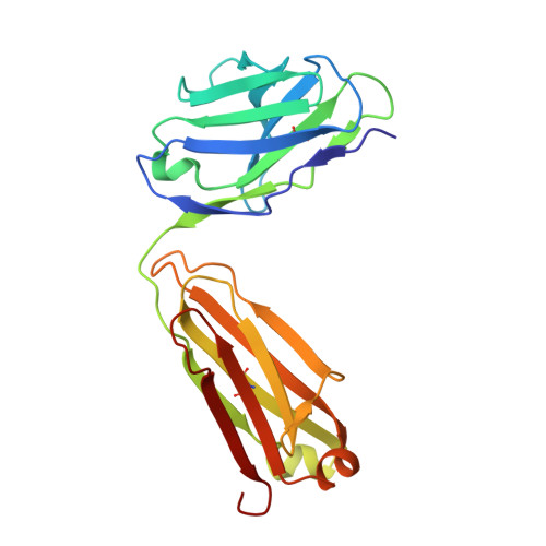
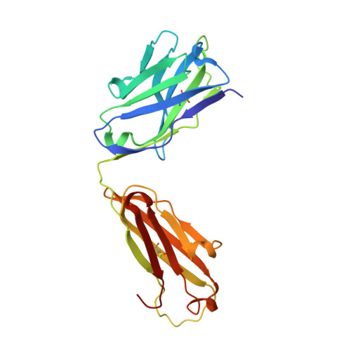
Domain Organization and Conformational Plasticity of the G Protein Effector, PDE6.

Zhang, Z.He, F.Constantine, R.Baker, M.L.Baehr, W.Schmid, M.F.Wensel, T.G.Agosto, M.A.

(2015) J Biol Chem 290: 12833-12843

  • DOI: https://doi.org/10.1074/jbc.M115.647636
  • Primary Citation of Related Structures:  
    3JAB, 3JBQ

  • PubMed Abstract: 

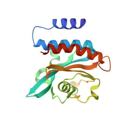
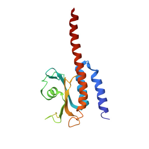
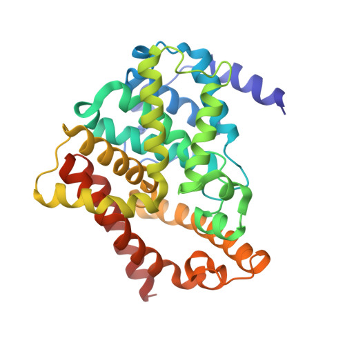
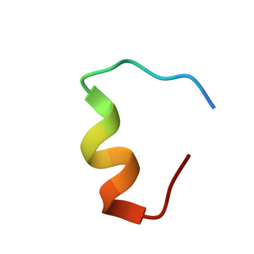
    The cGMP phosphodiesterase of rod photoreceptor cells, PDE6, is the key effector enzyme in phototransduction. Two large catalytic subunits, PDE6α and -β, each contain one catalytic domain and two non-catalytic GAF domains, whereas two small inhibitory PDE6γ subunits allow tight regulation by the G protein transducin. The structure of holo-PDE6 in complex with the ROS-1 antibody Fab fragment was determined by cryo-electron microscopy. The ∼11 Å map revealed previously unseen features of PDE6, and each domain was readily fit with high resolution structures. A structure of PDE6 in complex with prenyl-binding protein (PrBP/δ) indicated the location of the PDE6 C-terminal prenylations. Reconstructions of complexes with Fab fragments bound to N or C termini of PDE6γ revealed that PDE6γ stretches from the catalytic domain at one end of the holoenzyme to the GAF-A domain at the other. Removal of PDE6γ caused dramatic structural rearrangements, which were reversed upon its restoration.


  • Organizational Affiliation

    From the Department of Biochemistry and Molecular Biology, Baylor College of Medicine, Houston, Texas 77030 and.


Macromolecules
Find similar proteins by:  (by identity cutoff)  |  3D Structure
Entity ID: 1
MoleculeChains Sequence LengthOrganismDetailsImage
GafA domain of cone phosphodiesterase 6CA,
B [auth M]
180Bos taurusMutation(s): 0 
UniProt
Find proteins for A0A0M3KKT0 (Bos taurus)
Explore A0A0M3KKT0 
Go to UniProtKB:  A0A0M3KKT0
Entity Groups  
Sequence Clusters30% Identity50% Identity70% Identity90% Identity95% Identity100% Identity
UniProt GroupA0A0M3KKT0
Sequence Annotations
Expand
  • Reference Sequence
Find similar proteins by:  (by identity cutoff)  |  3D Structure
Entity ID: 2
MoleculeChains Sequence LengthOrganismDetailsImage
GafB domain of phosphodiesterase 2AC [auth B],
D [auth N]
185Bos taurusMutation(s): 0 
UniProt
Find proteins for P14099 (Bos taurus)
Explore P14099 
Go to UniProtKB:  P14099
Entity Groups  
Sequence Clusters30% Identity50% Identity70% Identity90% Identity95% Identity100% Identity
UniProt GroupP14099
Sequence Annotations
Expand
  • Reference Sequence
Find similar proteins by:  (by identity cutoff)  |  3D Structure
Entity ID: 3
MoleculeChains Sequence LengthOrganismDetailsImage
phosphodiesterase 5/6 chimera catalytic domainE [auth C],
G [auth O]
330Bos taurusMutation(s): 0 
UniProt
Find proteins for P16586 (Bos taurus)
Explore P16586 
Go to UniProtKB:  P16586
Find proteins for Q28156 (Bos taurus)
Explore Q28156 
Go to UniProtKB:  Q28156
Entity Groups  
Sequence Clusters30% Identity50% Identity70% Identity90% Identity95% Identity100% Identity
UniProt GroupsQ28156P16586
Sequence Annotations
Expand
  • Reference Sequence

Find similar proteins by:  Sequence   |   3D Structure  

Entity ID: 4
MoleculeChains Sequence LengthOrganismDetailsImage
phosphodiesterase 6 gamma subunit inhibitory peptideF [auth D],
H [auth P]
18Bos taurusMutation(s): 0 
UniProt
Find proteins for P04972 (Bos taurus)
Explore P04972 
Go to UniProtKB:  P04972
Entity Groups  
Sequence Clusters30% Identity50% Identity70% Identity90% Identity95% Identity100% Identity
UniProt GroupP04972
Sequence Annotations
Expand
  • Reference Sequence
Find similar proteins by:  (by identity cutoff)  |  3D Structure
Entity ID: 5
MoleculeChains Sequence LengthOrganismDetailsImage
IgG1-kappa 2E8 light chainI [auth L],
K [auth R]
214Mus musculusMutation(s): 0 
Entity Groups  
Sequence Clusters30% Identity50% Identity70% Identity90% Identity95% Identity100% Identity
Sequence Annotations
Expand
  • Reference Sequence
Find similar proteins by:  (by identity cutoff)  |  3D Structure
Entity ID: 6
MoleculeChains Sequence LengthOrganismDetailsImage
IgG1-kappa 2E8 heavy chainJ [auth H],
L [auth Q]
221Mus musculusMutation(s): 0 
Entity Groups  
Sequence Clusters30% Identity50% Identity70% Identity90% Identity95% Identity100% Identity
Sequence Annotations
Expand
  • Reference Sequence
Experimental Data & Validation

Experimental Data

  • Method: ELECTRON MICROSCOPY
  • Resolution: 11.0 Å
  • Aggregation State: PARTICLE 
  • Reconstruction Method: SINGLE PARTICLE 
EM Software:
TaskSoftware PackageVersion
RECONSTRUCTIONEMAN

Structure Validation

View Full Validation Report


Entry History 

Deposition Data

Revision History  (Full details and data files)

  • Version 1.0: 2015-06-10
    Type: Initial release
  • Version 1.1: 2018-07-18
    Changes: Data collection