3J8W

Cryo-EM reconstruction of quasi-HPV16 complex with H263.A2 Fab


Experimental Data Snapshot

  • Method: ELECTRON MICROSCOPY
  • Resolution: 13.0 Å
  • Aggregation State: PARTICLE 
  • Reconstruction Method: SINGLE PARTICLE 

wwPDB Validation   3D Report Full Report


This is version 1.2 of the entry. See complete history


Literature

Structural comparison of four different antibodies interacting with human papillomavirus 16 and mechanisms of neutralization.

Guan, J.Bywaters, S.M.Brendle, S.A.Lee, H.Ashley, R.E.Makhov, A.M.Conway, J.F.Christensen, N.D.Hafenstein, S.

(2015) Virology 483: 253-263

  • DOI: https://doi.org/10.1016/j.virol.2015.04.016
  • Primary Citation of Related Structures:  
    3J8V, 3J8W, 3J8Z

  • PubMed Abstract: 

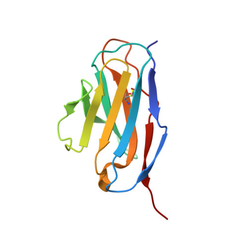
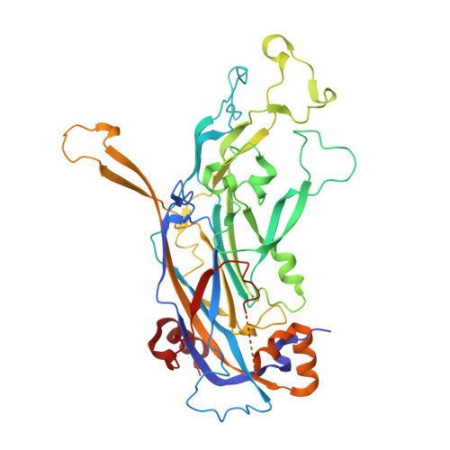
    Cryo-electron microscopy (cryo-EM) was used to solve the structures of human papillomavirus type 16 (HPV16) complexed with fragments of antibody (Fab) from three different neutralizing monoclonals (mAbs): H16.1A, H16.14J, and H263.A2. The structure-function analysis revealed predominantly monovalent binding of each Fab with capsid interactions that involved multiple loops from symmetry related copies of the major capsid protein. The residues identified in each Fab-virus interface map to a conformational groove on the surface of the capsomer. In addition to the known involvement of the FG and HI loops, the DE loop was also found to constitute the core of each epitope. Surprisingly, the epitope mapping also identified minor contributions by EF and BC loops. Complementary immunological assays included mAb and Fab neutralization. The specific binding characteristics of mAbs correlated with different neutralizing behaviors in pre- and post-attachment neutralization assays.


  • Organizational Affiliation

    Department of Medicine, The Pennsylvania State University College of Medicine, 500 University Drive, Hershey, PA 17033 USA.


Macromolecules
Find similar proteins by:  (by identity cutoff)  |  3D Structure
Entity ID: 1
MoleculeChains Sequence LengthOrganismDetailsImage
H263.A2 light chainA [auth L],
C [auth J],
E [auth K],
G [auth M]
109Mus musculusMutation(s): 0 
Entity Groups  
Sequence Clusters30% Identity50% Identity70% Identity90% Identity95% Identity100% Identity
Sequence Annotations
Expand
  • Reference Sequence
Find similar proteins by:  (by identity cutoff)  |  3D Structure
Entity ID: 2
MoleculeChains Sequence LengthOrganismDetailsImage
H263.A2 heavy chainB [auth H],
D [auth F],
F [auth G],
H [auth I]
118Mus musculusMutation(s): 0 
Entity Groups  
Sequence Clusters30% Identity50% Identity70% Identity90% Identity95% Identity100% Identity
Sequence Annotations
Expand
  • Reference Sequence
Find similar proteins by:  (by identity cutoff)  |  3D Structure
Entity ID: 3
MoleculeChains Sequence LengthOrganismDetailsImage
L1I [auth A],
J [auth B],
K [auth C],
L [auth D],
M [auth E]
455Human papillomavirus 16Mutation(s): 0 
UniProt
Find proteins for P03101 (Human papillomavirus type 16)
Explore P03101 
Go to UniProtKB:  P03101
Entity Groups  
Sequence Clusters30% Identity50% Identity70% Identity90% Identity95% Identity100% Identity
UniProt GroupP03101
Sequence Annotations
Expand
  • Reference Sequence
Experimental Data & Validation

Experimental Data

  • Method: ELECTRON MICROSCOPY
  • Resolution: 13.0 Å
  • Aggregation State: PARTICLE 
  • Reconstruction Method: SINGLE PARTICLE 
EM Software:
TaskSoftware PackageVersion
RECONSTRUCTIONAuto3DEM

Structure Validation

View Full Validation Report


Entry History 

Deposition Data

Revision History  (Full details and data files)

  • Version 1.0: 2015-05-06
    Type: Initial release
  • Version 1.1: 2015-06-03
    Changes: Database references
  • Version 1.2: 2018-07-18
    Changes: Data collection