3IQL

Crystal structure of the rat endophilin-A1 SH3 domain


Experimental Data Snapshot

  • Method: X-RAY DIFFRACTION
  • Resolution: 1.40 Å
  • R-Value Free: 0.154 
  • R-Value Work: 0.127 
  • R-Value Observed: 0.128 

wwPDB Validation   3D Report Full Report


This is version 1.2 of the entry. See complete history


Literature

SH3 domains from a subset of BAR proteins define a Ubl-binding domain and implicate parkin in synaptic ubiquitination.

Trempe, J.F.Chen, C.X.Grenier, K.Camacho, E.M.Kozlov, G.McPherson, P.S.Gehring, K.Fon, E.A.

(2009) Mol Cell 36: 1034-1047

  • DOI: https://doi.org/10.1016/j.molcel.2009.11.021
  • Primary Citation of Related Structures:  
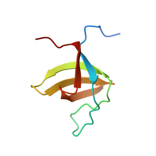
    2KNB, 3IQL

  • PubMed Abstract: 

    Mutations in the parkin gene are responsible for a common inherited form of Parkinson's disease (PD). Parkin is a RING-type E3 ubiquitin ligase with an N-terminal ubiquitin-like domain (Ubl). We report here that the parkin Ubl binds SH3 domains from endocytic BAR proteins such as endophilin-A with an affinity comparable to proline-rich domains (PRDs) from well-established SH3 partners. The NMR structure of the Ubl-SH3 complex identifies the PaRK extension, a unique C-terminal motif in the parkin Ubl required for SH3 binding and for parkin-mediated ubiquitination of endophilin-A in vitro. In nerve terminals, conditions that promote phosphorylation enhance the interaction between parkin and endophilin-A and increase the levels of ubiquitinated proteins within PRD-associated synaptic protein complexes in wild-type but not parkin knockout brain. The findings identify a pathway for the recruitment of synaptic substrates to parkin with the potential to explain the defects in synaptic transmission observed in recessive forms of PD.


  • Organizational Affiliation

    Department of Biochemistry, McGill University, Montréal, Québec, Canada.


Macromolecules
Find similar proteins by:  (by identity cutoff)  |  3D Structure
Entity ID: 1
MoleculeChains Sequence LengthOrganismDetailsImage
Endophilin-A1
A, B
71Rattus norvegicusMutation(s): 0 
Gene Names: Sh3d2aSh3gl2Sh3p4
UniProt
Find proteins for O35179 (Rattus norvegicus)
Explore O35179 
Go to UniProtKB:  O35179
Entity Groups  
Sequence Clusters30% Identity50% Identity70% Identity90% Identity95% Identity100% Identity
UniProt GroupO35179
Sequence Annotations
Expand
  • Reference Sequence
Experimental Data & Validation

Experimental Data

  • Method: X-RAY DIFFRACTION
  • Resolution: 1.40 Å
  • R-Value Free: 0.154 
  • R-Value Work: 0.127 
  • R-Value Observed: 0.128 
  • Space Group: C 1 2 1
Unit Cell:
Length ( Å )Angle ( ˚ )
a = 64.338α = 90
b = 34.156β = 107.79
c = 62.729γ = 90
Software Package:
Software NamePurpose
ADSCdata collection
MOLREPphasing
REFMACrefinement
HKL-2000data reduction
HKL-2000data scaling

Structure Validation

View Full Validation Report


Entry History 

Deposition Data

Revision History  (Full details and data files)

  • Version 1.0: 2009-11-10
    Type: Initial release
  • Version 1.1: 2011-07-13
    Changes: Version format compliance
  • Version 1.2: 2023-09-06
    Changes: Data collection, Database references, Derived calculations, Refinement description