3I5R

PI3K SH3 domain in complex with a peptide ligand


Experimental Data Snapshot

  • Method: X-RAY DIFFRACTION
  • Resolution: 1.70 Å
  • R-Value Free: 0.223 
  • R-Value Work: 0.182 
  • R-Value Observed: 0.184 

wwPDB Validation   3D Report Full Report


This is version 1.2 of the entry. See complete history


Literature

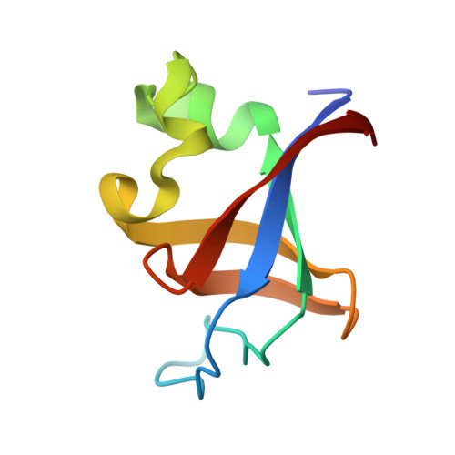
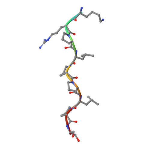
Structural studies of the phosphatidylinositol 3-kinase (PI3K) SH3 domain in complex with a peptide ligand: role of the anchor residue in ligand binding.

Batra-Safferling, R.Granzin, J.Modder, S.Hoffmann, S.Willbold, D.

(2010) Biol Chem 391: 33-42

  • DOI: https://doi.org/10.1515/BC.2010.003
  • Primary Citation of Related Structures:  
    3I5R, 3I5S

  • PubMed Abstract: 

    Src homology 3 (SH3) domains are mediators of protein-protein interactions. They comprise approximately 60 amino acid residues and are found in many intracellular signaling proteins. Here, we present the crystal structure of the SH3 domain from phosphatidylinositol 3-kinase (PI3K) in complex with the 12-residue proline-rich peptide PD1R (HSKRPLPPLPSL). The crystal structure of the PI3K SH3-PD1R complex at a resolution of 1.7 A reveals type I ligand orientation of the bound peptide with an extended conformation where the central portion forms a left-handed type II polyproline (PPII) helix. The overall structure of the SH3 domain shows minimal changes on ligand binding. In addition, we also attempted crystallization with another peptide ligand (PD1) where the residue at anchor position P(-3) is a tyrosine. The crystals obtained did not contain the PD1 ligand; instead, the ligand binding site is partially occupied by residues Arg18 and Trp55 from the symmetry-related PI3K SH3 molecule. Considering these crystal structures of PI3K SH3 together with published reports, we provide a comparative analysis of protein-ligand interactions that has helped us identify the individual residues which play an important role in defining target specificity.


  • Organizational Affiliation

    Institut für Strukturbiologie und Biophysik ISB-2, Forschungszentrum Jülich, D-52425 Jülich, Germany. r.batra-safferling@fz-juelich.de


Macromolecules
Find similar proteins by:  (by identity cutoff)  |  3D Structure
Entity ID: 1
MoleculeChains Sequence LengthOrganismDetailsImage
Phosphatidylinositol 3-kinase regulatory subunit alpha83Homo sapiensMutation(s): 0 
Gene Names: GRB1PIK3R1
UniProt & NIH Common Fund Data Resources
Find proteins for P27986 (Homo sapiens)
Explore P27986 
Go to UniProtKB:  P27986
PHAROS:  P27986
GTEx:  ENSG00000145675 
Entity Groups  
Sequence Clusters30% Identity50% Identity70% Identity90% Identity95% Identity100% Identity
UniProt GroupP27986
Sequence Annotations
Expand
  • Reference Sequence

Find similar proteins by:  Sequence   |   3D Structure  

Entity ID: 2
MoleculeChains Sequence LengthOrganismDetailsImage
Peptide ligand12N/AMutation(s): 0 
Entity Groups  
Sequence Clusters30% Identity50% Identity70% Identity90% Identity95% Identity100% Identity
Sequence Annotations
Expand
  • Reference Sequence
Experimental Data & Validation

Experimental Data

  • Method: X-RAY DIFFRACTION
  • Resolution: 1.70 Å
  • R-Value Free: 0.223 
  • R-Value Work: 0.182 
  • R-Value Observed: 0.184 
  • Space Group: P 31 2 1
Unit Cell:
Length ( Å )Angle ( ˚ )
a = 60.365α = 90
b = 60.365β = 90
c = 46.525γ = 120
Software Package:
Software NamePurpose
ADSCdata collection
MOLREPphasing
PHENIXrefinement
MOSFLMdata reduction
SCALAdata scaling

Structure Validation

View Full Validation Report


Entry History 

Deposition Data

Revision History  (Full details and data files)

  • Version 1.0: 2010-03-02
    Type: Initial release
  • Version 1.1: 2011-07-13
    Changes: Version format compliance
  • Version 1.2: 2023-09-06
    Changes: Data collection, Database references, Refinement description