3HYR

Structural Insight into G Protein Coupling and Regulation of Fe2+ Membrane Transport


Experimental Data Snapshot

  • Method: X-RAY DIFFRACTION
  • Resolution: 2.20 Å
  • R-Value Free: 0.287 
  • R-Value Work: 0.225 
  • R-Value Observed: 0.228 

wwPDB Validation   3D Report Full Report


This is version 1.2 of the entry. See complete history


Literature

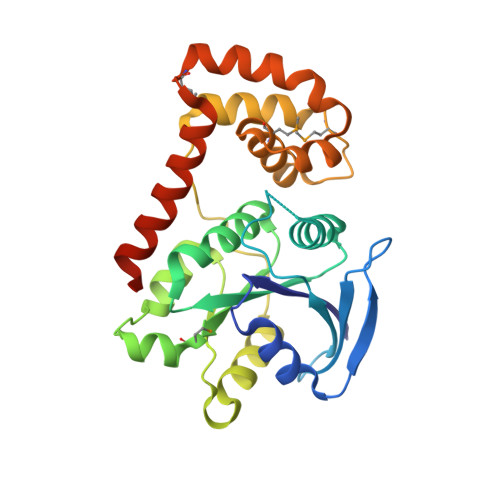
Structural basis of GDP release and gating in G protein coupled Fe2+ transport.

Guilfoyle, A.Maher, M.J.Rapp, M.Clarke, R.Harrop, S.Jormakka, M.

(2009) EMBO J 28: 2677-2685

  • DOI: https://doi.org/10.1038/emboj.2009.208
  • Primary Citation of Related Structures:  
    3HYR, 3HYT

  • PubMed Abstract: 

    G proteins are key molecular switches in the regulation of membrane protein function and signal transduction. The prokaryotic membrane protein FeoB is involved in G protein coupled Fe(2+) transport, and is unique in that the G protein is directly tethered to the membrane domain. Here, we report the structure of the soluble domain of FeoB, including the G protein domain, and its assembly into an unexpected trimer. Comparisons between nucleotide free and liganded structures reveal the closed and open state of a central cytoplasmic pore, respectively. In addition, these data provide the first observation of a conformational switch in the nucleotide-binding G5 motif, defining the structural basis for GDP release. From these results, structural parallels are drawn to eukaryotic G protein coupled membrane processes.


  • Organizational Affiliation

    Centenary Institute, Sydney, New South Wales, Australia.


Macromolecules
Find similar proteins by:  (by identity cutoff)  |  3D Structure
Entity ID: 1
MoleculeChains Sequence LengthOrganismDetailsImage
Ferrous iron transport protein B
A, B, C
270Escherichia coliMutation(s): 0 
Gene Names: feoBb3409JW3372
UniProt
Find proteins for P33650 (Escherichia coli (strain K12))
Explore P33650 
Go to UniProtKB:  P33650
Entity Groups  
Sequence Clusters30% Identity50% Identity70% Identity90% Identity95% Identity100% Identity
UniProt GroupP33650
Sequence Annotations
Expand
  • Reference Sequence
Small Molecules
Modified Residues  1 Unique
IDChains TypeFormula2D DiagramParent
MSE
Query on MSE
A, B, C
L-PEPTIDE LINKINGC5 H11 N O2 SeMET
Experimental Data & Validation

Experimental Data

  • Method: X-RAY DIFFRACTION
  • Resolution: 2.20 Å
  • R-Value Free: 0.287 
  • R-Value Work: 0.225 
  • R-Value Observed: 0.228 
  • Space Group: C 1 2 1
Unit Cell:
Length ( Å )Angle ( ˚ )
a = 146.12α = 90
b = 84.44β = 108.46
c = 66.24γ = 90
Software Package:
Software NamePurpose
SHARPphasing
REFMACrefinement
MOSFLMdata reduction
SCALAdata scaling

Structure Validation

View Full Validation Report


Entry History 

Deposition Data

Revision History  (Full details and data files)

  • Version 1.0: 2009-08-25
    Type: Initial release
  • Version 1.1: 2011-07-13
    Changes: Advisory, Version format compliance
  • Version 1.2: 2013-11-13
    Changes: Database references