3HF2

Crystal structure of the I401P mutant of cytochrome P450 BM3


Experimental Data Snapshot

  • Method: X-RAY DIFFRACTION
  • Resolution: 2.20 Å
  • R-Value Free: 0.245 
  • R-Value Work: 0.186 
  • R-Value Observed: 0.189 

wwPDB Validation   3D Report Full Report


Ligand Structure Quality Assessment 


This is version 1.3 of the entry. See complete history


Literature

A Highly Active Single-Mutation Variant of P450(BM3) (CYP102A1)

Whitehouse, C.J.C.Bell, S.G.Yang, W.Yorke, J.A.Blanford, C.F.Strong, A.J.F.Morse, E.J.Bartlam, M.Rao, Z.Wong, L.-L.

(2009) Chembiochem 10: 1654-1656

  • DOI: https://doi.org/10.1002/cbic.200900279
  • Primary Citation of Related Structures:  
    3HF2

  • PubMed Abstract: 

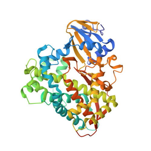
    The power of proline: Bold amino acid substitutions in sensitive protein regions are frequently unproductive, while more subtle mutations can be sufficient to bring about dramatic changes. But introducing proline at the residue next to the sulfur ligand in P450(BM3) (CYP102A1) has the unexpected and desirable effect of enhancing the activity of this fatty acid hydroxylase with a broad range of non-natural substrates, as illustrated by the figure.


  • Organizational Affiliation

    Department of Chemistry, University of Oxford, Inorganic Chemistry Laboratory, South Parks Road, Oxford, UK.


Macromolecules
Find similar proteins by:  (by identity cutoff)  |  3D Structure
Entity ID: 1
MoleculeChains Sequence LengthOrganismDetailsImage
Bifunctional P-450/NADPH-P450 reductase
A, B
482Priestia megateriumMutation(s): 1 
Gene Names: P450BM-3
EC: 1.14.14.1
UniProt
Find proteins for P14779 (Priestia megaterium (strain ATCC 14581 / DSM 32 / CCUG 1817 / JCM 2506 / NBRC 15308 / NCIMB 9376 / NCTC 10342 / NRRL B-14308 / VKM B-512 / Ford 19))
Explore P14779 
Go to UniProtKB:  P14779
Entity Groups  
Sequence Clusters30% Identity50% Identity70% Identity90% Identity95% Identity100% Identity
UniProt GroupP14779
Sequence Annotations
Expand
  • Reference Sequence
Small Molecules
Ligands 1 Unique
IDChains Name / Formula / InChI Key2D Diagram3D Interactions
HEM
Query on HEM

Download Ideal Coordinates CCD File 
C [auth A],
D [auth B]
PROTOPORPHYRIN IX CONTAINING FE
C34 H32 Fe N4 O4
KABFMIBPWCXCRK-RGGAHWMASA-L
Experimental Data & Validation

Experimental Data

  • Method: X-RAY DIFFRACTION
  • Resolution: 2.20 Å
  • R-Value Free: 0.245 
  • R-Value Work: 0.186 
  • R-Value Observed: 0.189 
  • Space Group: P 1 21 1
Unit Cell:
Length ( Å )Angle ( ˚ )
a = 58.67α = 90
b = 145.843β = 97.33
c = 63.229γ = 90
Software Package:
Software NamePurpose
HKL-2000data collection
PHASERphasing
REFMACrefinement
HKL-2000data reduction
HKL-2000data scaling

Structure Validation

View Full Validation Report


Ligand Structure Quality Assessment 


Entry History 

Deposition Data

Revision History  (Full details and data files)

  • Version 1.0: 2009-06-30
    Type: Initial release
  • Version 1.1: 2011-07-13
    Changes: Advisory, Version format compliance
  • Version 1.2: 2021-11-10
    Changes: Database references, Derived calculations
  • Version 1.3: 2023-11-01
    Changes: Data collection, Refinement description