3GSY

Structure of berberine bridge enzyme in complex with dehydroscoulerine


Experimental Data Snapshot

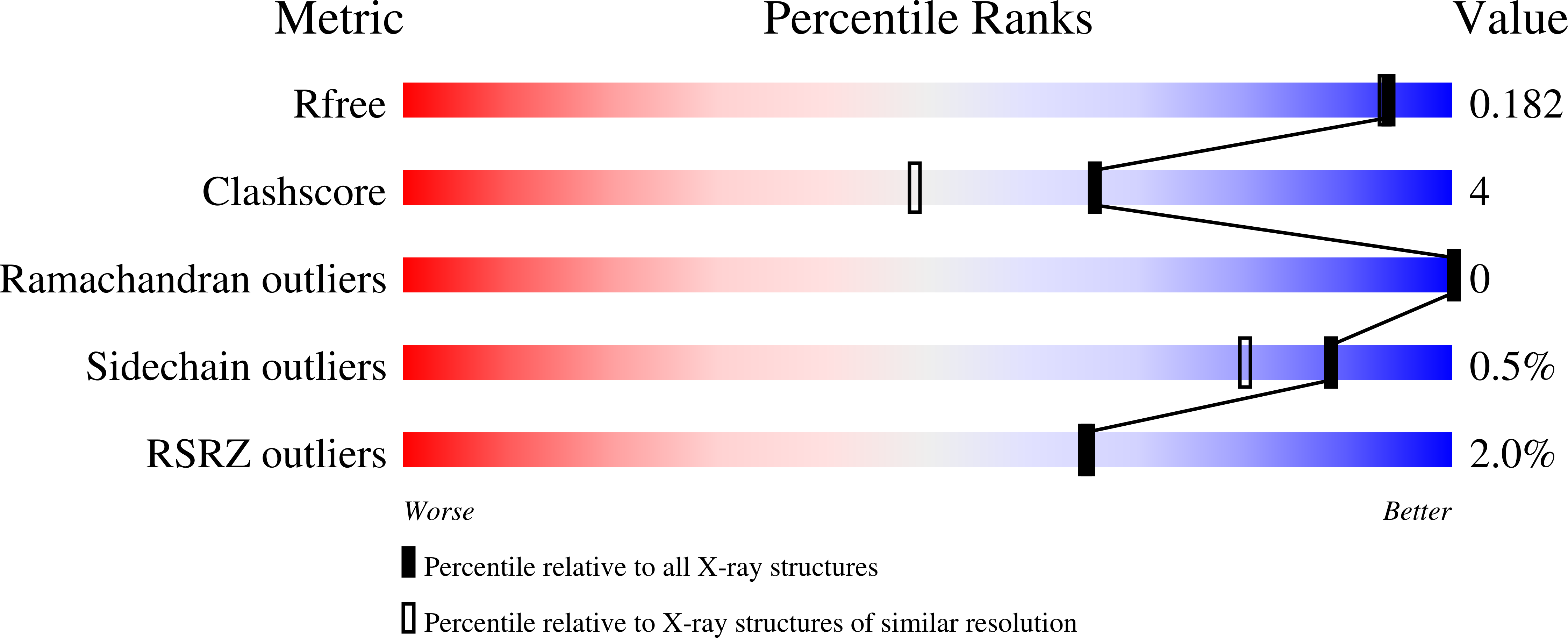
  • Method: X-RAY DIFFRACTION
  • Resolution: 1.63 Å
  • R-Value Free: 0.185 
  • R-Value Work: 0.162 
  • R-Value Observed: 0.163 

wwPDB Validation   3D Report Full Report


Ligand Structure Quality Assessment 


This is version 2.1 of the entry. See complete history


Literature

Berberine bridge enzyme catalyzes the six electron oxidation of (S)-reticuline to dehydroscoulerine.

Winkler, A.Puhl, M.Weber, H.Kutchan, T.M.Gruber, K.Macheroux, P.

(2009) Phytochemistry 70: 1092-1097

  • DOI: https://doi.org/10.1016/j.phytochem.2009.06.005
  • Primary Citation of Related Structures:  
    3GSY

  • PubMed Abstract: 

    Berberine bridge enzyme catalyzes the stereospecific oxidation and carbon-carbon bond formation of (S)-reticuline to (S)-scoulerine. In addition to this type of reactivity the enzyme can further oxidize (S)-scoulerine to the deeply red protoberberine alkaloid dehydroscoulerine albeit with a much lower rate of conversion. In the course of the four electron oxidation, no dihydroprotoberberine species intermediate was detectable suggesting that the second oxidation step leading to aromatization proceeds at a much faster rate. Performing the reaction in the presence of oxygen and under anoxic conditions did not affect the kinetics of the overall reaction suggesting no strict requirement for oxygen in the oxidation of the unstable dihydroprotoberberine intermediate. In addition to the kinetic characterization of this reaction we also present a structure of the enzyme in complex with the fully oxidized product. Combined with information available for the binding modes of (S)-reticuline and (S)-scoulerine a possible mechanism for the additional oxidation is presented. This is compared to previous reports of enzymes ((S)-tetrahydroprotoberberine oxidase and canadine oxidase) showing a similar type of reactivity in different plant species.


  • Organizational Affiliation

    Institute of Biochemistry, Graz University of Technology, Petersgasse 12/II, 8010 Graz, Austria.


Macromolecules
Find similar proteins by:  (by identity cutoff)  |  3D Structure
Entity ID: 1
MoleculeChains Sequence LengthOrganismDetailsImage
Reticuline oxidase; Berberine bridge-forming enzyme519Eschscholzia californicaMutation(s): 0 
Gene Names: BBE1
EC: 1.21.3.3
UniProt
Find proteins for P30986 (Eschscholzia californica)
Explore P30986 
Go to UniProtKB:  P30986
Entity Groups  
Sequence Clusters30% Identity50% Identity70% Identity90% Identity95% Identity100% Identity
UniProt GroupP30986
Sequence Annotations
Expand
  • Reference Sequence
Oligosaccharides

Help

Entity ID: 2
MoleculeChains Length2D Diagram Glycosylation3D Interactions
beta-D-mannopyranose-(1-4)-2-acetamido-2-deoxy-beta-D-glucopyranose-(1-4)-2-acetamido-2-deoxy-beta-D-glucopyranose
B
3N-Glycosylation
Glycosylation Resources
GlyTouCan:  G15407YE
GlyCosmos:  G15407YE
GlyGen:  G15407YE
Small Molecules
Ligands 4 Unique
IDChains Name / Formula / InChI Key2D Diagram3D Interactions
FAD
Query on FAD

Download Ideal Coordinates CCD File 
C [auth A]FLAVIN-ADENINE DINUCLEOTIDE
C27 H33 N9 O15 P2
VWWQXMAJTJZDQX-UYBVJOGSSA-N
DEH
Query on DEH

Download Ideal Coordinates CCD File 
D [auth A]2,9-dihydroxy-3,10-dimethoxy-5,6-dihydroisoquino[3,2-a]isoquinolinium
C19 H18 N O4
DRAPVRZDBZHUPL-UHFFFAOYSA-O
NAG
Query on NAG

Download Ideal Coordinates CCD File 
E [auth A]2-acetamido-2-deoxy-beta-D-glucopyranose
C8 H15 N O6
OVRNDRQMDRJTHS-FMDGEEDCSA-N
MG
Query on MG

Download Ideal Coordinates CCD File 
F [auth A]MAGNESIUM ION
Mg
JLVVSXFLKOJNIY-UHFFFAOYSA-N
Experimental Data & Validation

Experimental Data

  • Method: X-RAY DIFFRACTION
  • Resolution: 1.63 Å
  • R-Value Free: 0.185 
  • R-Value Work: 0.162 
  • R-Value Observed: 0.163 
  • Space Group: P 41 21 2
Unit Cell:
Length ( Å )Angle ( ˚ )
a = 68.94α = 90
b = 68.94β = 90
c = 247.26γ = 90
Software Package:
Software NamePurpose
XSCALEdata scaling
PHENIXrefinement
PDB_EXTRACTdata extraction
PHENIXphasing

Structure Validation

View Full Validation Report


Ligand Structure Quality Assessment 


Entry History 

Deposition Data

Revision History  (Full details and data files)

  • Version 1.0: 2009-06-30
    Type: Initial release
  • Version 1.1: 2011-07-13
    Changes: Non-polymer description, Version format compliance
  • Version 2.0: 2020-07-29
    Type: Remediation
    Reason: Carbohydrate remediation
    Changes: Atomic model, Data collection, Database references, Derived calculations, Structure summary
  • Version 2.1: 2023-09-06
    Changes: Data collection, Database references, Refinement description, Structure summary