3GPL

Crystal structure of the ternary complex of RecD2 with DNA and ADPNP


Experimental Data Snapshot

  • Method: X-RAY DIFFRACTION
  • Resolution: 2.50 Å
  • R-Value Free: 0.273 
  • R-Value Work: 0.232 
  • R-Value Observed: 0.232 

wwPDB Validation   3D Report Full Report


Ligand Structure Quality Assessment 


This is version 1.2 of the entry. See complete history


Literature

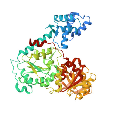
Mechanistic basis of 5'-3' translocation in SF1B helicases.

Saikrishnan, K.Powell, B.Cook, N.J.Webb, M.R.Wigley, D.B.

(2009) Cell 137: 849-859

  • DOI: https://doi.org/10.1016/j.cell.2009.03.036
  • Primary Citation of Related Structures:  
    3GP8, 3GPL

  • PubMed Abstract: 

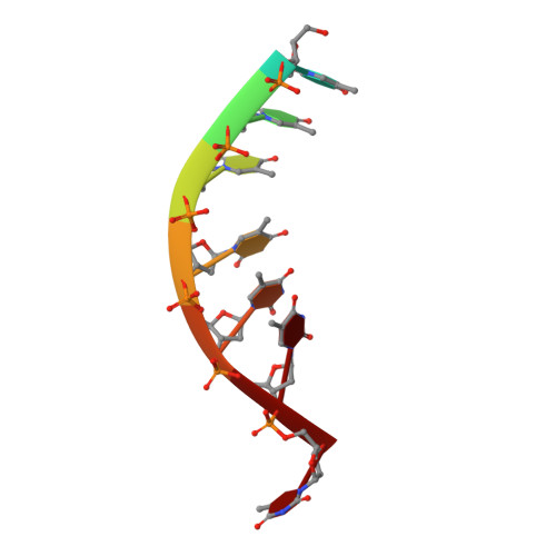
    Superfamily 1B (SF1B) helicases translocate in a 5'-3' direction and are required for a range of cellular activities across all domains of life. However, structural analyses to date have focused on how SF1A helicases achieve 3'-5' movement along nucleic acids. We present crystal structures of the complex between the SF1B helicase RecD2 from Deinococcus radiodurans and ssDNA in the presence and absence of an ATP analog. These snapshots of the reaction pathway reveal a nucleotide binding-induced conformational change of the two motor domains that is broadly reminiscent of changes observed in other SF1 and SF2 helicases. Together with biochemical data, the structures point to a step size for translocation of one base per ATP hydrolyzed. Moreover, the structures also reveal a mechanism for nucleic acid translocation in the 5'-3' direction by SF1B helicases that is surprisingly different from that of 3'-5' translocation by SF1A enzymes, and explains the molecular basis of directionality.


  • Organizational Affiliation

    Cancer Research UK Clare Hall Laboratories, The London Research Institute, Blanche Lane, South Mimms, Potters Bar, Herts EN63LD, UK.


Macromolecules

Find similar proteins by:  (by identity cutoff)  |  3D Structure
Entity ID: 1
MoleculeChains Sequence LengthOrganismDetailsImage
Exodeoxyribonuclease V, subunit RecD, putative
A, B
574Deinococcus radiodurans R1 = ATCC 13939 = DSM 20539Mutation(s): 0 
Gene Names: DR_1902recD2
UniProt
Find proteins for Q9RT63 (Deinococcus radiodurans (strain ATCC 13939 / DSM 20539 / JCM 16871 / CCUG 27074 / LMG 4051 / NBRC 15346 / NCIMB 9279 / VKM B-1422 / R1))
Explore Q9RT63 
Go to UniProtKB:  Q9RT63
Entity Groups  
Sequence Clusters30% Identity50% Identity70% Identity90% Identity95% Identity100% Identity
UniProt GroupQ9RT63
Sequence Annotations
Expand
  • Reference Sequence

Find similar nucleic acids by:  Sequence   |   3D Structure  

Entity ID: 2
MoleculeChains LengthOrganismImage
5'-D(*T*TP*TP*TP*TP*TP*TP*T)-3'C [auth X],
D [auth Y]
8N/A
Sequence Annotations
Expand
  • Reference Sequence
Experimental Data & Validation

Experimental Data

  • Method: X-RAY DIFFRACTION
  • Resolution: 2.50 Å
  • R-Value Free: 0.273 
  • R-Value Work: 0.232 
  • R-Value Observed: 0.232 
  • Space Group: P 1 21 1
Unit Cell:
Length ( Å )Angle ( ˚ )
a = 66.12α = 90
b = 130.36β = 107.34
c = 80.05γ = 90
Software Package:
Software NamePurpose
MAR345data collection
PHASERphasing
CNSrefinement
XDSdata reduction
XDSdata scaling

Structure Validation

View Full Validation Report


Ligand Structure Quality Assessment 


Entry History 

Deposition Data

Revision History  (Full details and data files)

  • Version 1.0: 2009-06-16
    Type: Initial release
  • Version 1.1: 2011-07-13
    Changes: Version format compliance
  • Version 1.2: 2023-09-06
    Changes: Data collection, Database references, Derived calculations, Refinement description